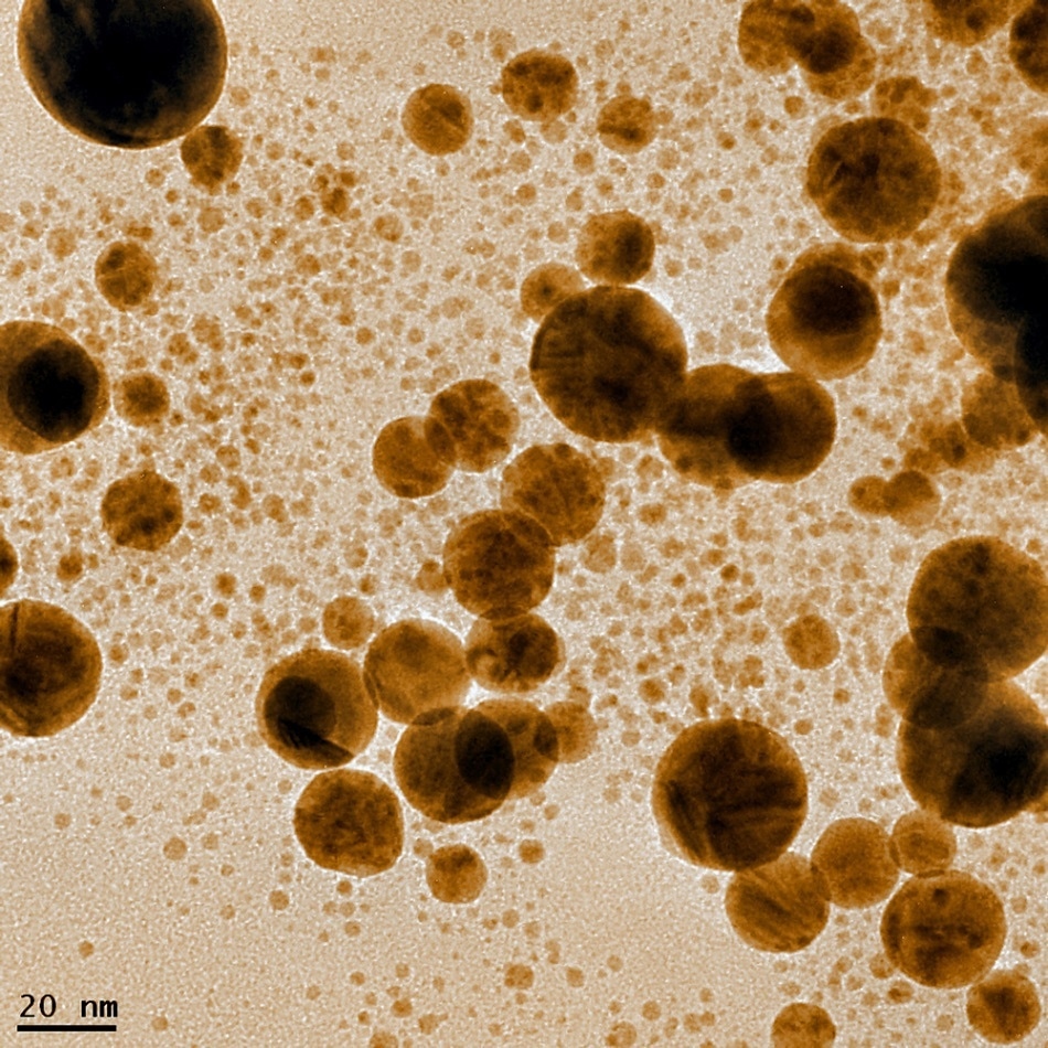
Researchers from the University of Graz’s Institute of Physics and Institute for Electron Microscopy and Nanoanalysis have recently demonstrated a new method for 3D reconstruction of plasmonic nearfields. Ulrich Hohenester and Gerald Kothleitner’s approach has been found suitable to reconstruct the 3D plasmonic topographic imaging of larger nanoparticles and is found to be applicable to dielectic or metallic nanostructures of various complexities and sizes.

As light consists of particles called photons, conducting media such as plasmas and metals contain plasmons, a quantum of plasma oscillations. Plasmons are rapid oscillations of electronic fluid in metals1.
The study of the interaction between electromagnetic field and the free electrons in metals that are excited by the electrical component of the light is called plasmonics1. By using light to create electronic oscillations, or plasmons, in the metallic nanoparticles, the field of plasmonics, due to its ability to confine light at the nanoscale, shows promise for a wide variety of applications. However, the direct observations of these plasmonic nearfields by utilizing optical methods has been difficult due to the diffraction limit of the light.
Scanning transmission electron microscopy (STEM) equipped with electron energy loss spectroscopy (EELS) presents an alternative approach to detect these plasmonic oscillations. When the surface of metal nanoparticles is focused with a beam of swift electrons, a small amount of the electrons’ kinetic energy is lost due to the excitation of particle plasmons. By rapidly scanning the surface of the specimen, the energy loss can be analyzed, and the plasmonic nearfields can be mapped with a nanometer spatial and sub-eV resolution.
Although this method has been extensively used to study particle plasmons, interpreting EELS signal has a few shortcomings2. Firstly, valuable information along the beam propagation direction is masked because the EELS determines the plasmonic nearfields by measuring the energy loss across the specimen. Secondly, it is hard to correlate sample quantity to the EELS signal. Lastly, only in special cases would the EELS be able to probe the photonic local density of states (LDOS), a measure used to determine the states occupied by the photon.
Based on the assumptions that the probe-sample interactions are local in space, three dimensional (3D) tomographic imaging utilizes a series of projections from different viewing angles to create a three-dimensional image.

 Subscribe to our Nanotechnology Newsletters
The tomographic scheme utilizing the EELS is based on the assumptions that the nanoparticles are much smaller than the wavelength of light, and the plasmonic response is directed by a single resonance. While this method has been successfully used in earlier work conducted on silver nanocubes, this method cannot be applied to larger nanoparticles beyond the quasistatic approximation and this method also requires that the light does not penetrate the nanoparticle2.
Therefore, a more robust and reliable method that can provide 3D tomographic imaging could be beneficial to the fields of nanophotonics and plasmonics.
Ulrich Hohenester and Gerald Kothleitner’s team first performed tomographic experiments by measuring the EELS spectrum images (SIs) and high angle annular dark field (HAADF) images for different tilt angles. The HAADF data is then used for particle geometry, which is used for computing generic eigenmode basis (Ek)2.
The green tensor is decomposed into the eigenmodes computed is used to create reprojected EELS maps based on the expansion coefficients determined by the minimization procedure. In the third step, the photonic environment of plasmonic nanoparticles is visualized based on the best expansion coefficients and the quantities of interest such as plasmonic LDOS are finally computed.
Even though the simulated and experimental EELS spectra is not perfect, the reprojected EELS maps are in accordance with the measured ones. This research team is currently working on determining the applicability of their method for composite structures, as well as investigating the coupling effects between plasmonic nanoparticles and quantum emmiters such as quantum dots. The method developed in this study has the potential to be applied to a variety of nanophotonic systems such as dielectric spheres.
References:
- “Plasmonics” – Stanford University
- “Tomographic imaging of the photonic environment of plasmonic nanoparticles A. Horl, G. Haberfehlner, et al. Nature Communications. (2017) DOI: 10.1038/s41467-017-00051-3.
Image Credit:
Georgy Shafeev/ Shutterstock.com
Disclaimer: The views expressed here are those of the author expressed in their private capacity and do not necessarily represent the views of AZoM.com Limited T/A AZoNetwork the owner and operator of this website. This disclaimer forms part of the Terms and conditions of use of this website.