 By JanakyReviewed by Lexie CornerUpdated on Jul 24 2024
Cancer remains a significant global health challenge, with nearly 20 million new cases and approximately 9.7 million deaths in 2022.1 Early detection and accurate assessment of tumor margins are crucial for improving survival rates and treatment success.2
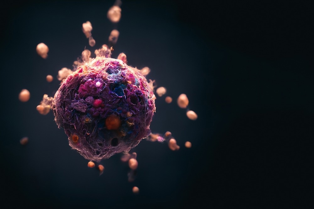
Image Credits: CI Photos/Shutterstock.com
Delayed or inaccessible cancer care reduces survival chances, increases treatment complications, and raises healthcare costs, underscoring the need for timely diagnosis and accessible healthcare systems worldwide.
Nanotechnology can enhance cancer diagnosis and treatment in multiple ways. Utilizing nanoparticles (NPs) and quantum dots (QDs) to detect cancer at the molecular level is a key area of research.3-5
Scanning and Imaging Cancerous Tumors
Key imaging techniques used in cancer research and clinical settings include magnetic resonance imaging (MRI), computed tomography (CT), positron emission tomography (PET), and ultrasound imaging (US).6 These techniques have improved cancer diagnosis but have limitations, especially in detecting early-stage tumors.
For instance, MRI offers high spatial resolution but suffers from long imaging times and low sensitivity.6 CT provides detailed images based on X-ray attenuation but poses a radiation risk and is less effective at detecting small tumors.6
NPs can be used to improve cancer cell visibility by binding to specific receptors on the cell surface, generating strong signals detectable via imaging instruments.7 For example, gold NPs can be utilized in CT scans to provide high-contrast images due to their surface plasmon resonance properties.8
Multifunctional NPs can be further engineered by modifying their shells or surfaces or attaching affinity ligands to target specific cancer cells. This targeted imaging approach can be applied using various imaging techniques, including MRI, PET, and CT.7
First-generation NPs have already been designed, developed, and tested in living systems, showing promise for future cancer imaging and therapy applications.
Discover More: Properties and Applications of Gold Nanoparticles
Detecting Cancer by Analyzing Tissue Samples
Fluorescent immunoassays (FIA) are promising for early cancer diagnosis.9 These assays use fluorescent tags attached to antibodies that bind to cancer biomarkers, emitting a measurable signal.
Nanotechnology enhances FIAs by allowing the detection of very low biomarker levels, typical in early-stage cancer.9 The incorporation of NPs also facilitates the detection of multiple biomarkers simultaneously.10
Nanostructures also induce metal-enhanced fluorescence, where light interacts with metallic nanostructures' electromagnetic fields, significantly enhancing fluorescence-based detection sensitivity.11
Nanomaterials like the "disk-coupled dots-on-pillar antenna array" (D2PA) amplify light from fluorescent labels, enhancing detection sensitivity up to 7,400-fold for proteins like Immunoglobulin G.11 This amplification, due to highly confined electromagnetic fields in "hotspot" regions, is crucial for early disease diagnosis.
Quantum dots (QDs), semiconductor particles at the nanoscale, possess excellent light-emitting properties that are useful in medical imaging. Graphene-based QDs are highly effective in biosensors, identifying cancer biomarkers like antigens, enzymes, hormones, and proteins.12 The enhanced sensitivity and specificity of QD-based sensors make them valuable tools in diagnosing various cancers early and monitoring treatment effectiveness.
Enhancing Cancer Immunotherapy with NPs
One major advancement in cancer immunotherapy is the development of NP vaccines. These vaccines deliver tumor-specific antigens directly to immune cells, like dendritic cells, initiating strong T-cell responses.13
Once activated, T cells destroy infected or cancerous cells, making T cell responses vital for cancer therapy. NPs help attract and activate T cells in the tumor environment, improving the efficacy of treatments like immune checkpoint blockade and adoptive T cell transfer.13
By improving the delivery and presentation of these antigens, NP vaccines can elicit a potent and precise immune response against cancer cells. This capability to influence the immune system helps create a more hostile environment for cancer cells, aiding treatment.13
Additionally, these NPs can be engineered with immunostimulatory properties to boost the immune response further, addressing issues of poor immunogenicity and off-target effects.
NPs can also be utilized in combined treatments, incorporating photothermal therapy, photodynamic therapy, and gene therapy.14 For instance, in photothermal therapy, NPs absorb radiation and convert it into heat to kill cancer cells; in photodynamic therapy, they produce reactive oxygen species to damage cancer cells.14
These combined approaches enhance the overall effectiveness of cancer treatments, reduce side effects, and improve targeting, offering a comprehensive strategy to combat cancer.
Challenges and Future Outlook
Nanotechnology presents promising advancements for cancer treatment and diagnosis but faces significant challenges in implementation. Designing effective nanomedicines tailored to specific cancer types remains complex and costly, hindering scalability from laboratory settings to clinical applications.15
Regulatory challenges like FDA approval processes must keep pace with technological progress. Safety concerns require rigorous assessment to mitigate potential toxicological risks and ensure environmental sustainability.15 Manufacturing nanomedicines at scale while maintaining quality and affordability is also challenging.
Looking forward, advanced NPs are being developed for targeted drug delivery, improving effectiveness and reducing toxicity. Advances in personalized medicine with nanotechnology and machine learning can tailor treatments based on individual patient profiles.15
The multimodal imaging capabilities of NPs promise improved tumor localization and monitoring. Combination therapies using nanocarriers may also help combat drug resistance and enhance treatment outcomes.
Future research will prioritize sustainability, developing eco-friendly nanomaterials and manufacturing practices to reduce environmental impact.
More from AZoNano: Quantum Dot Labeling: Precision and Sensitivity in Biological Research
References and Further Reading
- National Cancer Institute. (2024). Cancer Statistics. [Online] National Cancer Institute. Available at: https://www.cancer.gov/about-cancer/understanding/statistics
- World Health Organization. (2024). Promoting Cancer Early Diagnosis. [Online] World Health Organization. Available at: https://www.who.int/activities/promoting-cancer-early-diagnosis
- Nasir, A., Khan, A., Li, J., Naeem, M., Khalil, AA., Khan, K., Qasim, M. (2021). Nanotechnology: A tool for diagnostics and treatment of cancer. Current topics in medicinal chemistry. DOI: 10.2174/1568026621666210701144124
- Grodzinski, P., Silver, M., Molnar, LK. (2006). Nanotechnology for cancer diagnostics: promises and challenges. Expert review of molecular diagnostics. DOI: 10.1586/14737159.6.3.307
- Alrushaid, N., Khan, FA., Al-Suhaimi, EA., Elaissari, A. (2023). Nanotechnology in cancer diagnosis and treatment. Pharmaceutics. DOI: 10.3390/pharmaceutics15031025
- Luengo Morato, Y., Ovejero Paredes, K., Lozano Chamizo, L., Marciello, M., Filice, M. (2021). Recent advances in multimodal molecular imaging of cancer mediated by hybrid magnetic nanoparticles. Polymers. DOI: 10.3390/polym13172989
- Barreto, J. A., O’Malley, W., Kubeil, M., Graham, B., Stephan, H., Spiccia, L. (2011). Nanomaterials: applications in cancer imaging and therapy. Advanced materials. DOI: 10.1002/adma.201100140
- Xi, D., Dong, S., Meng, X., Lu, Q., Meng, L., Ye, J. (2012). Gold nanoparticles as computerized tomography (CT) contrast agents. Rsc Advances. DOI: 10.1039/C2RA21263C
- Devi, RV., Doble, M., Verma, RS. (2015). Nanomaterials for early detection of cancer biomarker with special emphasis on gold nanoparticles in immunoassays/sensors. Biosensors and Bioelectronics. DOI: 10.1016/j.bios.2015.01.066
- Barbosa, AI., Rebelo, R., Reis, RL., Bhattacharya, M., Correlo, VM. (2021). Current nanotechnology advances in diagnostic biosensors. Medical Devices & Sensors. DOI: 10.1002/mds3.10156
- Badshah, M. A., Koh, N. Y., Zia, A. W., Abbas, N., Zahra, Z., & Saleem, M. W. (2020). Recent developments in plasmonic nanostructures for metal enhanced fluorescence-based biosensing. Nanomaterials. DOI: 2079-4991/10/9/1749
- Iannazzo, D., Espro, C., Celesti, C., Ferlazzo, A., Neri, G. (2021). Smart biosensors for cancer diagnosis based on graphene quantum dots. Cancers. DOI: 2072-6694/13/13/3194
- Fan, Y., Moon, JJ. (2015). Nanoparticle drug delivery systems designed to improve cancer vaccines and immunotherapy. Vaccines. DOI: 2076-393X/3/3/662
- Nam, J., Son, S., Park, KS., Zou, W., Shea, LD., Moon, JJ. (2019). Cancer nanomedicine for combination cancer immunotherapy. Nature Reviews Materials. DOI: 10.1038/s41578-019-0108-1
- Kemp, JA., Kwon, YJ. (2021). Cancer nanotechnology: current status and perspectives. Nano convergence. DOI: 10.1186/s40580-021-00282-7
Disclaimer: The views expressed here are those of the author expressed in their private capacity and do not necessarily represent the views of AZoM.com Limited T/A AZoNetwork the owner and operator of this website. This disclaimer forms part of the Terms and conditions of use of this website.