Jun 8 2009
Canadian Light Source (CLS) staff scientist Luca Quaroni and Dr. Alan Casson, Head of the Department of Surgery at the University of Saskatchewan (U of S) used the synchrotron's infrared microscope to identify tissue afflicted with a condition known as Barrett's Esophagus from chemical fingerprints associated with the disease, which can lead to esophageal cancer.
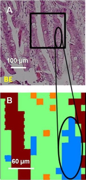 Visible light micrograph of Barrett's esophagus tissue (A) and a synchrotron infrared image (B) from the Canadian Light Source. The light blue area corresponds to an area rich in glycoproteins, a biomarker for Barrett's esophagus. (Credit: Image courtesy of Luca Quaroni, CLS)
"The advantage to using microscopes with synchrotron light is that it allows us to identify chemical biomarkers inside specific cells," says Dr. Quaroni, who conducted the synchrotron analysis. "Often the differences between healthy and malignant tissue can be quite small, but the differences seen here were quite striking. This is a good proof of concept for developing a traceable technique that matches what can be seen at the macroscopic scale using microscopic samples."
The team analyzed preserved samples of healthy and diseased tissue that Dr. Casson had collected during esophageal biopsies. Using a technique known as Fourier Transform Infrared Microscopy, Quaroni and Casson identified specific chemicals – known as biomarkers - within the individual cells that make up the tissue. It was found that increased concentrations of particular biomarkers such as glycoproteins were associated with the Barrett's tissue.
Barrett's Esophagus (BE) occurs when the cells that normally line the esophagus – the tube that connects our throat to our stomach – are replaced by cells that resemble those that line the intestine. While BE only affects approximately one percent of Americans, the number of people diagnosed with the condition is on the rise, with the increasing incidence of chronic heartburn (gastro-esophageal reflux disease or GERD) considered a risk factor for developing BE. The disease in turn can lead to an aggressive form of cancer known as esophageal adenocarcinoma.
Currently, diagnosing BE depends on the skill and experience of individual pathologists examining biopsy samples from patients, often relying on subjective criteria. Identifying biomarkers that can be associated with a particular condition provides an additional tool for diagnosis.
The finding is published in the June, 2009 issue of the Royal Society of Chemistry journal, The Analyst.