The first Carl Zeiss AURIGA™ CrossBeam® electron/ion microscope was officially put into service today at the Ludwig-Maximilians University (LMU) in Munich. There is considerable global interest in this system, which was introduced only four months ago. A number of orders have already been placed; the demo systems are reserved weeks in advance. AURIGA™ combines a high-resolution electron microscope with an ion beam which enables precise incisions in the specimens to be examined, thus providing access to structures below the specimen surface. In addition to specimen imaging on a nano scale, a huge variety of detectors also permit a chemical-physical analysis of the specimen.
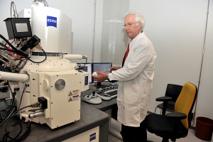 Professor Gerhard Wanner, head of the ultra-structure research work group at the Ludwig-Maxiliams University in Munich presents the worldwide first AURIGA™ CrossBeam® Workstation. At the Biocenter of the LMU the AURIGA will be used for a wide range of applications, including the manufacture of high-resolution cross-section images of cell nuclei.
Professor Gerhard Wanner, head of the ultra-structure research work group at the university is extremely happy to use the instrument: “A major field in the Biocenter will be the 3D reconstruction of tissues, cells and their sub-structures. The versatility of this system offers a series of completely new examination possibilities which we hope will lead to important insights into ultra-structural and functional interrelationships.”
Requests for the AURIGA system have been received from institutes and industry around the world: “The flexibility and versatility of the system are obviously key factors contributing to this interest. Bio/life science, materials research, the semiconductor industry, solar cell research – we are receiving request from practically all important fields of application,” explains Product Manager Dr. Daniel Kraft.