John Spence, a physicist at Arizona State University, is a longtime user of the Advanced Light Source at Lawrence Berkeley National Laboratory, where he has contributed to major advances in lensless imaging. It's a particularly apt propensity for someone who works with x-rays, since they can't be focused with ordinary lenses.
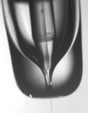 In the gas dynamic virtual nozzle, liquid flows through a internal capillary, issuing some distance from the opening in the outer tube through which the gas flows. Approaching the narrow opening, gas pressure and speed increase, focusing the thin stream of liquid until it is so small only a single protein or virus can fit into each droplet. Credit: courtesy John Spence, ASU
As new light sources evolve to produce brighter x-rays in faster pulses, lensless imaging becomes ever more critical for science. Among the promises of superbright, ultrafast x-ray pulses is the ability to solve the structure of the complicated molecules from which our bodies are made. All living things are made of proteins and nucleic acids, but relatively few of the atomic structures of the thousands, perhaps millions, of varieties of proteins are known.
The Linac Coherent Light Source (LCLS) will soon begin operation at the SLAC National Accelerator Laboratory in Palo Alto, California, using energetic electrons from a linear accelerator to produce coherent x-rays with an instrument called a free electron laser (FEL). The x-rays will be delivered 120 times a second in pulses only a tenth of a trillionth of a second long – about the time it takes light to travel the width of a human hair. These brief, bright pulses offer a novel approach to the problem of protein structure.
Unfolding the origami
Proteins begin as strings of amino acids that fold themselves into an amazing variety of origami-like structures, whose bumps and crannies and distribution of electrical charges determine how they act individually or fit together to form complex molecular machines. Simple organisms like viruses often consist of a few proteins fitted together to enclose a thread of DNA or RNA.
Proteins are usually large molecules containing many thousands of atoms. Drug molecules are much smaller, and do their work by attaching themselves to the larger protein molecules. A knowledge of the arrangement of a protein's atoms is therefore a great help to drug designers, who like to understand how a drug molecule will dock with a protein to promote or inhibit its activity, or cripple the organism of which it is a part.
Until now, the best way to solve the structure of a protein or virus has been with x-ray crystallography. The crystal consists of many copies of the protein or virus arranged in regular order. As the crystal rotates in the x-ray beam, x-rays scatter off the atoms and reveal – once these complex diffraction patterns have been converted into a 3-D image by computers – how the electrons, and thus the atoms, are arranged.
But many proteins can't be crystallized at all, and others are so difficult to crystallize it's virtually impossible to obtain crystals large enough to use in today's light sources.
Ultrafast, ultrabright x-rays offer a way past this dilemma. The idea is that a quick pulse of tightly focused x-rays can be diffracted from a microcrystal or even a single protein or virus in solution. The pulse is so brief that it comes and goes before any of the atoms can move, freezing their orientation like a strobe light. Just as important, a sufficiently brief pulse may terminate before radiation damage effects can start. In this way it can outrun radiation damage, always one of the fundamental limitations to imaging in biology.
Another quick pulse could be diffracted from another copy of the protein in a different orientation. As the process is repeated, diffractions from different angles give the overlapping views needed for the computer to construct a 3-D image of the structure.
It's a great idea, but as Spence notes, there are a few problems. "So as not to scatter, the x-ray beam has to be in a high vacuum, but a protein or virus in its natural state is usually wet. As in T. S. Eliot's Wasteland, water is life. How do we maintain the protein or virus in an aqueous environment inside the vacuum?"
Click here to read the full press release