People often have strong opinions on the "right" firmness of mattresses for themselves, and, as it turns out, some cell types have similar preferences for their support structures. Now a research team from the National Institute of Standards and Technology (NIST) and the National Institutes of Health (NIH) has developed a way to offer cells a three-dimensional scaffold that varies over a broad range of degrees of stiffness to determine where they develop best.
Their recently published technique* is a way to rapidly optimize 3D cell growth media to meet the developmental needs of specific cell types for a wide variety of potential tissue-replacement therapies.
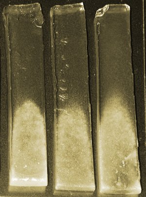 Experimental hydrogel scaffolds prepared at NIST for culturing bone cells clearly show the dependence of bone mineralization on the stiffness of the gel. Deposited minerals are denser at the bottoms of the gradient gels, which are progressively stiffer from top to bottom. Strips are approx. 1 X 6 cm. (Color added for emphasis.) Credit: Chatterjee, NIST
Tissue engineering is a relatively new field that is developing methods to grow or regenerate bodily tissues—skin, bone, cartilage, blood vessels, perhaps one day even whole organs—to replace those damaged by injury or disease. One of the key challenges in the field is developing appropriate three-dimensional “scaffolds,” artificial materials that can hold tissue progenitor cells and allow them to be nurtured and supported while they multiply and develop into desired tissues. Research has shown that cells often need to develop in a 3D environment if they are to mature and differentiate properly.
Hydrogels—most familiar for their use in soft contact lenses—are a promising material for tissue scaffolds. They consist of a loose network of polymer chains that is swollen with water; in fact, like the majority of the body’s tissues, they are mostly water.
But, says NIST materials scientist Kaushik Chatterjee, deciding on a hydrogel is just the beginning. “Now you’ve got these gels, what sort of properties do you want? What gets you the best kind of whatever tissue you’re after—in our case, bone? We focused on stiffness because cells are known to sense and respond to changes in the stiffness of their environment.”
To test this, the research team developed a method to create samples of a typical hydrogel used in biomedical research, PEGDM**, where the stiffness of the gel increases smoothly from one end of the sample to the other. This approach, using smoothly varying gradients of compounds to test many possible combinations simultaneously, is called combinatorial screening. NIST has pioneered such techniques for a variety of materials problems***, but this research is one of the first applications of combinatorial screening to 3D scaffolds for tissue engineering. The team tested the technique on mouse osteoblasts—cells responsible for building bone—mixed in with the PEGDM gel. Interestingly, although cell survival rates were higher at the softer end of the test strips and got progressively worse towards the stiffer ends, cell differentiation and mineralization, which are measures of how well the cells actually develop into bone tissue, did the reverse. Fewer cells survive in a stiff gel, but those that do are much more active in building bone. That result, of course, is specific to osteoblasts, says Chatterjee, “These are bone cells and they seem to like the stiffer environment more than softer ones, but you could apply something similar to, say, nerve cells, and they might like the softer ones more.”
In addition, the researchers note, the gel stiffness gradient induced a matching gradient in the tissue mineralization. This is potentially important, they say, because tissue gradients often occur naturally at the interfaces of, for example, teeth or ligaments, so 3D scaffold gradients could be a valuable tool for engineering graded tissues for regenerative medicine.
The research was supported by NIST and NIH.