A heart formed of polymers, a tuberculosis bacterium camouflaged as an exotic blossom and a dragon made of a magnesium-titanium compound: it is often hard to tell whether the entries to the Carl Zeiss Nano Image Contest are artistically structured paintings or photos taken under the microscope.
More than 70 researchers from around the globe are currently presenting their fascinating nano masterpieces to the general public in an online contest. Many visitors to the website have already used the opportunity to vote for their favorite image.
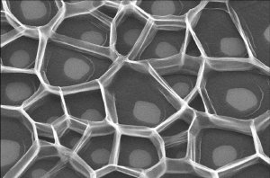 Combs of nano tubes: The current voting record is held by Peter Nirmalraj with his Scanning Electron Microscope photo.
To date, not only classical black-and-white photos but also several stained microscope images have been submitted. Most of the images received until now belong to the category of scanning electron microscopy (SEM), while fewer have been entered in the categories of transmission electron microscopy (TEM) and helium ion microscopy (HIM). Therefore, more photos from these areas would be particularly welcome. Peter Nirmalraj from Trinity College Dublin is the current leader in the overall competition.
All users of ZEISS particle beam systems can submit their most beautiful photos to the Nano Image Contest before 29 August. The website is available for voting for another two weeks until 12 September in order to give voters the opportunity to upload images not submitted until the latter part of the submission period. The winners of the four categories will each receive a pair of cinemizer Plus video glasses from 12 September Carl Zeiss.
Source: Carl Zeiss