Oct 6 2010
Researchers from Oxford have recently presented exciting new data applying nanoparticle tracking analysis (NTA) from Nanosight to size and count both cellular microvesicles and exosomes at a low concentration and, when used in conjunction with fluorescent labels, to selectively determine and analyse specific types of vesicle within a complex sample.
This took place during a two-day conference in Oxford, “Micro and Nanovesicles in Health and Disease”, organised by Dr Paul Harrison from the Oxford Haemophilia & Thrombosis Centre at the Churchill Hospital in Oxford and Ian Sargent, Professor of Reproductive Science in the Nuffield Department of Obstetrics and Gynaecology, University of Oxford.
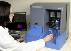 The NanoSight NS500 system like the one being used in Oxford by the Sargent group
Most researchers concur that the high levels of microvesicles and/or exosomes are associated with (i.e. potential biomarkers for) thrombotic diseases, cardiovascular disease and some cancers. Leading the research at the Nuffield Department of Obstetrics and Gynaecology, University of Oxford, Professor Ian Sargent says “many cells shed small vesicles in a regulated way which plays a key role in intercellular communication. In general, there are two types of vesicle: microvesicles (100nm – 1 µm in diameter) which directly bud from the plasma membrane and nanovesicles (exosomes 30nm - 100nm) which are released by exocytosis from multivesicular bodies of the endosome. Both are involved in cell signalling. They carry diverse membrane and cytosolic proteins as well as messenger and microRNAs. They can affect the physiology of their target cells in various ways, from inducing intracellular signalling following binding to receptors, to conferring new properties after the acquisition of new receptors, enzymes or genetic material by fusion or endocytosis. They participate in physiological processes including haemostasis and thrombosis, inflammation, immune interactions and angiogenesis.
Continuing, Sargent said: “NanoSight’s NTA technique is a major step forward in analytical capability taking the limits of flow cytometry down almost an order of magnitude. It is rapid and, in common with flow cytometry, characterizes polydispersity well.”
Also at this conference Edwin van der Pol from the Academic Medical Center at the University of Amsterdam presented a theoretical comparison of analytical techniques for microvesicles and exosomes. This confirmed the advantages of using NTA for studying vesicles sized from 50-400nm. The previously preferred technique was flow cytometry. However, van der Pol concluded this has a practical lower limit of 300nm. Similarly electrozone sensing is not able detect at such small sizes. He added that while Dynamic Light Scattering (DLS) is able to identify very small particles, it generally biases towards large particles in polydisperse samples so this mis-reporting renders it of little value. It is also not able to make concentration measurement.
Jeremy Warren, NanoSight CEO, commented “The work of this important group is a significant milestone for us. It is the first step toward directing NanoSight’s capability as a platform for biomarker detection”.
In the closing session, Dr Karl Morten, also from University of Oxford, described NanoSight’s useful role in rapidly assessing newly developed nanoparticles, as he summarised a range of roles of nanotechnology in drug delivery.
This conference bought together 135 delegates in this rapidly growing area of interest. There were five groups from the UK, Netherlands and USA who have recently added NanoSight’s NTA to their characterisation capability.