Aug 13 2013
NanoSight reports on how Nanoparticle Tracking Analysis, NTA, is being used at St George's, University of London, to explore extracellular vesicles as a potential source of biomarkers, by identifying proteins or peptides differentially expressed between healthy subjects and patients with rare inherited diseases
St George's, University of London, is the UK's only independent medical and healthcare higher education institution. Dr Bridget Bax is a senior research fellow in the Clinical Sciences Division where the main focus of her group's research is to improve the understanding of the pathogenic mechanisms of rare inherited diseases and to develop novel therapies for translation into the clinical setting. One of their major areas of interest is the identification and validation of biomarkers in patients with the rare and fatal disease mitochondrial neurogastrointestinal encephalomyopathy (MNGIE).
They are currently exploring the use of extracellular vesicles as source of biomarkers by identifying proteins or peptides differentially expressed between healthy subjects and patients with MNGIE. The goals of this work are three-fold: to understand the pathophysiological mechanisms and metabolic derangements observed in patients with MNGIE; to provide a means of monitoring more effectively clinical and biochemical response to therapy; and to enable the tracking of disease progression in diagnosed patients.
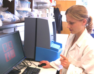
Describing her work, Dr Bax said "We have used several methods in the lab to isolate both exosomes and microparticles. We needed a reproducible method that would allow us to i) quantitate and size the extracellular vesicles we isolated and ii) determine whether the isolation technique employed affected these parameters. Nanoparticle Tracking Analysis has allowed us to quantify extracellular vesicles with diameters in the range of 50 to 1000 nm. This was particularly important for us because we specifically wanted to study exosomes which have a diameter ranging from 40 to 100 nm."
Prior to using NTA, initially the group used electron microscopy and fluorescence-activated cell sorting (FACS), a biophysical technique used in flow cytometry. Using FACS, Dr Bax said "We were unable to detect exosomes and found that a significant number of microparticles were missed due to not detecting vesicles with a diameter <300 nm. Although it was possible to size extracellular vesicles using electron microscopy, this is a time intensive technique and has the potential disadvantages of causing shrinkage."
Dr Bax went on to discuss her thoughts on using NTA: "The benefits are the ability to detect particles within the size range of interest. NTA uses small sample volumes which is extremely important in terms of the rare diseases we study. We find the system easy to use."
To find out about the company and to learn more about particle characterization using NanoSight's unique Nanoparticle Tracking Analysis solutions, visit www.nanosight.com and register to receive the next issue of NanoTrail, the company's electronic newsletter.