Think of it as an emergency room exam for cells. In the span of a few minutes, scientists at Lawrence Berkeley National Laboratory can produce three-dimensional CAT scans of entire cells, revealing their internal structure down to the smallest organelle.
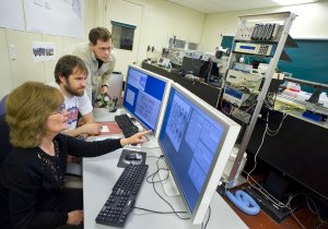 Carolyn Larabell (left), the director of the National Center for X-ray Tomography, leads a team of scientists who are developing soft X-ray tomography as a new tool for visualizing the internal architecture of whole cells.
The new imaging technology could speed up a wide range of research that requires scientists to track changes in a cell's internal structure, from analyzing how a pharmaceutical prevents a microbe from causing yeast infections to learning the best way to squeeze biofuel from algae.
The images are courtesy of the National Center for X-ray Tomography, which is located at Berkeley Lab's Advanced Light Source. At its heart is a soft X-ray microscope that can image cells at a resolution of about 50 nanometers. Soft X-rays are so-named because they are less penetrating than those used to diagnose a broken bone - perfect for microscopic samples.
The microscope uses the intense light generated by the Advanced Light Source to take two-dimensional X-ray images of cells, and then digitally stitches these slices into three-dimensional images of whole cells. It is the only soft X-ray tomography facility dedicated to biochemical and biomedical research.
Recently, a team of scientists from Berkeley Lab, Stanford University, and the University of California, San Francisco used the facility to capture the changes that occur when Candida albicans is exposed to a new and promising antifungal therapy.
C. albicans is a single-cell microbe that lives on the skin and in the genitourinary and gastrointestinal tracts of people. It is found in approximately 80 percent of humans and is usually harmless, but it sometimes becomes pathogenic and causes yeast infections. This transformation from good to bad is marked by the growth of a long filament, called a hypha, which extends from the microbe like a tail.
The soft X-ray tomography images revealed that microbes treated with the candidate drug molecules did not develop long hyphae, meaning the therapy successfully blocked C. albicans from becoming infectious. The images also uncovered a spike in the number and size of lipid bodies inside C. albicans cells, as well as newly formed holes in the cells' nucleolus, which is a structure in the nucleus.
Some of these changes would have taken months of work to view using other microscopy techniques, and a few would have been missed altogether.
"But this only took us a few days using soft x-ray tomography," says Gerry McDermott , a guest scientist in Berkeley Lab's Physical Biosciences Division and a scientist at the University of California, San Francisco.
"We saw cellular changes that hadn't been seen in the past. Because of its high resolution and very little sample prep time, this new technology may help expedite drug discovery," says McDermott.
Electron microscopy is currently the workhorse of cellular imaging. It offers incredibly high-resolution snapshots of cellular architecture. But it's slow: a sample slated for analysis must undergo weeks of preparation. Another common tool is fluorescence microscopy. But it only images molecules within a cell that are specially tagged, not the entire cell.
"Our findings would have been very difficult to obtain using other techniques," adds Maho Uchida of Berkeley Lab's Physical Biosciences Division and the University of California, San Francisco. "The tail of C. albicans is 50 microns long. It would have taken many electron microscopy images and months of work to see it - but we got it in one sweep."
In addition to learning how to fight disease, scientists can train the facility's high-throughput and high-resolution imaging capabilities on many scientific challenges, including the hunt for carbon-neutral sources of energy.
"We can use soft X-ray tomography to measure and quantify the formation of biofuel droplets in algae cells," says McDermott.
Scientists from the National Center for X-ray Tomography will also soon add high-resolution cryogenic light microscopy to the facility's existing X-ray tomography' toolkit. When combined with soft X-ray microscopy, the facility will be able to produce images that show both a cell's structure and the location of chemically labeled molecules or proteins within the cell.