Jun 1 2010
To diagnose cancer reliably, doctors usually conduct a biopsy including tissue analysis - which is a time-consuming process. A microscopic image sensor, fitted in an endoscope, is being developed for in vivo cancer diagnosis, to speed up the detection of tumors.
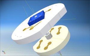 The fiber transmits the laser light to the microscanner mirror. Both are fitted in the tip of the endoscope. (© Fraunhofer IPMS)
Early detection is the key to the successful treatment of cancer. But not every lump turns out to be a malignant tumor. To find out whether cancerous cells are present, doctors usually conduct a biopsy and examine the removed tissue under the microscope. This process is not only very stressful for the patient but also highly time consuming. Research scientists at the Fraunhofer Institute for Photonic Microsystems IPMS in Dresden are aiming to considerably speed up cancer diagnosis. They have developed a microscope head with a diameter of just eight millimeters which can optically resolve and magnify tissue cells measuring just 10 to 20 micrometers. Fitted in the tip of an endoscope it will be used for in vivo cancer diagnosis, inserted in the body as in a minimally invasive surgical operation. The scientists envision that the MEMS (micro-electro-mechanical system) microscope head will eliminate the need for biopsies. Diagnosis in real time would enable doctors to decide on the necessary course of treatment more quickly.
"Microscopic image recorders that can be used on endoscopes have not been available up to now. We have developed the first laser-based sensor for this purpose," says Dr. Michael Scholles, business unit manager at the IPMS. "In classic endoscopy using macroscopic imaging, the job can be done by CCD or CMOS image sensors, as used in digital cameras and cellphones. For endomicroscopy, however, MEMS-based image sensors are highly advantageous because they can magnify even the smallest object fields, such as cells, without the need for a large lens. We have combined the sensor with a microscanner mirror to achieve the required resolution of 10 micrometers and can therefore massively magnify the tiniest structures."
But how does the system function? The laser itself is located in the operating theater. The laser light is conducted via a transmitting fiber to the microscanner mirror fitted in the tip of the endoscope. This deflects the laser beam and illuminates the suspicious tissue specifically. A glass-fiber bundle in the tip of the endoscope transmits the reflected light to the external sensor, which thus receives a signal containing the image information. A detector precisely measures the position of the scanner mirror, indicating which area of the scene is being illuminated at the specific point in time. A two-dimensional image can thus be completely reconstructed by combining the position and image sensor signals.
"An important aspect of the development was to produce a suitable microassembly for the endoscope head. Here we faced the challenge of making the complete system suitable for installation in the endoscope, and we managed to do it. In future our microscope head will be produced in large quantities in an automated process for subsequent installation in endoscopes," explains Scholles. The expert envisages a wide range of applications for the system: "It could be used not only in medical and biological microscopy but also in technical endoscopy, for instance to examine cavities in buildings or to inspect the insides of engines and turbines." The microscope head has already been produced as a demonstrator and can be seen at the Optatec trade show in Frankfurt from June 15 to 18 (Hall 3, Stand D50).