Aug 11 2010
In a development that holds potential for both data storage and biomedical imaging, Ohio State University researchers have used a new technique to obtain the highest-ever resolution MRI scan of the inside of a magnet.
Chris Hammel, Ohio Eminent Scholar in Experimental Physics, and his colleagues took a tiny magnetic disk -- measuring only 2 micrometers (millionths of a meter) across and 40 nanometers (billionths of a meter) thick -- and were able to obtain magnetic resonance images its interior.
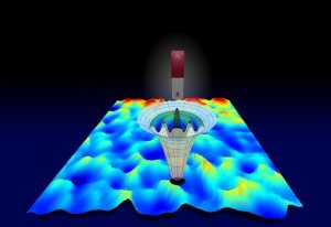 Researchers at Ohio State University have developed a new type of magnetic resonance that can see inside magnetic materials. Here, slight variations in the structure of a thin magnetic film are evident as variations in ferromagnetic resonance frequency, represented by changes in color. Above the film is a representation of a polarized magnetic tip that scans the material. (Credit: Image courtesy of Ohio State University.)
The resulting image -- with each "pixel" one tenth the size of the disk itself -- is the highest-resolution image ever taken of the magnetic fields and interactions inside of a magnet.
Why look inside magnets? Because studying the material's behavior at these tiny scales is key to incorporating them into computer chips and other electronic devices.
The researchers report their findings in the August 12 issue of the journal Nature.
In 2008, Hammel's team debuted a new kind of high-resolution scanning system that combines three different kinds of technology: MRI, ferromagnetic resonance, and atomic force microscopy.
Ferromagnets -- the type of magnet used in this study -- are magnets made of ferrous metal such as iron. Common household refrigerator magnets are ferromagnets.
Because ferromagnets retain a particular polarization once magnetized, they are already essential components in today's computers and other electronics, where they provide data storage alongside computer chips. But smaller magnets built directly into a computer chip could do even more, Hammel explained.
"We know that shrinking these magnets to the nanoscale and building them directly inside electronics would enable these devices to do more, and with less power consumption," Hammel said. "But a key barrier has always been the difficulty of imaging and characterizing nanomagnets."
Typical MRI machines work by inducing a magnetic field inside non-magnetic objects, such as the body. Since ferromagnets are already magnetic, conventional MRI can't see inside them.
The combination technique that the Ohio State researchers invented is called "scanned probe ferromagnetic resonance imaging," or scanned probe FMRI, and it involves detecting a magnetic signal using a tiny silicon bar with an even tinier magnetic probe on its tip.
In Nature, they report a successful demonstration of the technique, as they imaged the inside of the magnetic disk 0.2 micrometers (200 nanometers) at a time. They used a thin film of a commercially available nickel-iron magnetic alloy called Permalloy for the disk.
"In essence, we were able to conduct ferromagnetic resonance measurements on a small fraction of the disk, then move our probe over a little bit and do magnetic resonance there, and so on," explained Denis Pelekhov, director of the ENCOMM NanoSystems Laboratory at Ohio State. "Using these results, we could see how the magnetic properties vary inside the disk."
Experts suspect that computer chips equipped with tiny magnets might one day provide high-density data storage. Computers with magnets in their central processing units (CPUs) would never have to boot up. The entire computer would be contained inside the CPU, making such devices even smaller and less power-hungry as well.
Hammel believes that the technique could one day be useful tool in biomedical research labs. Researchers could use it to study tissue samples of the plaques that form in brain tissues and arteries, and perhaps develop better ways of detecting them in the body. Knowing how these plaques form could advance studies of many diseases, including Alzheimer's and atherosclerosis.
Hammel and Pelekhov's co-authors on the paper include Inhee Lee, Yuri Obukhov, Gang Xiang,, Adam Hauser, Fengyuan Yang, and Palash Banerjee, all of the Department of Physics at Ohio State.
This research was funded by the Department of Energy.