Aug 12 2010
A new laser-equipped microscope at IU Bloomington's Light Microscopy Imaging Center makes it possible to examine biological samples with unprecedented detail in three dimensions.
The $1.2 million DeltaVision OMX super-resolution microscope from Applied Precision (Issaquah, Wash.) was paid for entirely with funds from the American Recovery and Reinvestment Act of 2009, through a National Institutes of Health program that supports high-end instrumentation at America's most deserving centers of higher education.
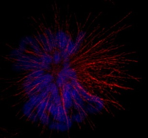 A fixed PTK (marsupial kidney cell line) cell in mitosis. The condensed chromosomes are stained with Hoechst stain and are shown in blue, while microtubules are labelled with an antibody to tubulin and are shown in red. Imaging was done in the structured illumination mode. More than 2000 images were taken and processed to create this image.
"It's a fantastic and unique acquisition for our university," said cell biologist Claire Walczak, the Imaging Center's executive director. "This super-resolution microscope, one of only 16 in the world and one of only 8 commercial units, is part of our vision to bring state-of-the-art technology to IU's life science researchers, to enable them to address questions that they did not have the ability to ask previously, due to the lack of appropriate technologies."
Walczak is a professor of biochemistry and molecular biology in the Medical Sciences Program Bloomington, an arm of the IU School of Medicine. Walczak also holds an adjunct appointment in the IU Bloomington Department of Biology and is part of the Biochemistry Program.
The imager is exceptionally fast in collecting images of a biological specimen, and this speed enables scientists to gather crucial data. The device uses laser light of four different colors to illuminate samples, while four extremely sensitive digital cameras capture images every 10 milliseconds at the imager's speediest setting. The device can produce as many as 5,000 full-color images per minute for its major task of producing high-resolution images. Known as a "structured illumination" microscope, the device will help IU scientists attain a better understanding of how proteins are distributed inside cells with unprecedented resolution.
Most high-technology light microscopes reach the limits of resolution at 250-300 nanometers -- the diameter of a small bacterial cell. The new OMX microscope IU has acquired can produce clear images down to 100 nanometers in the lateral dimension. Resolution along the z-axis (perpendicular, or coming out of the page) is somewhat lower but still tremendously improved relative to previous technologies.
"We'd envisioned this device would be most useful for microbiologists, cell biologists, and neurobiologists at IU," Walczak said. "But we expect scientists from many other fields will come up with creative ways to take advantage of it."
Light Microscopy Imaging Center (LMIC) Manager Jim Powers is responsible for training IU researchers -- as well as visitors -- to use the device.
"The Imaging Center is a user-oriented resource," Powers said. "Scientists rent time on our devices, and receive training to use them, but after that, we expect they'll be able to work independently."
IU scientists get a reduced rate when using the LMIC's many microscopes, due to the generous support from OVPR, the College of Arts and Sciences, Medical Sciences, and Optometry. At present, the OMX is still in a training mode in which Powers is working closely with Sid Shaw, an assistant professor of biology and the technical director of the LMIC, as well as IU research staff to calibrate the device and establish protocols for future, similar uses. The LMIC staff expects the instrument to be available to all IU researchers by September.
The arrival of the DeltaVision OMX microscope has spurred Walczak, Shaw, and Powers to consider LMIC's future needs. Partly because of the DeltaVision OMX's size, the LMIC is now out of physical space. In addition, the device produces so much data (4,000 images takes up about 1.5 gigabytes of hard drive space), Walczak and Powers said one of the center's next priorities is to improve the center's information technology infrastructure through continued collaboration with IU's Information Technology Services. Walczak and Powers want to ensure that the large data sets produced by the OMX imager can be stored rapidly -- as well as protected from power outages and other catastrophes.
Source: http://www.iub.edu/