Electron microscopes are among the most widely used scientific and medical tools for studying and understanding a wide range of materials, from biological tissue to miniature magnetic devices, at tiny levels of detail.
Now, researchers at the National Institute of Standards and Technology (NIST) have found a novel and potentially widely applicable method to expand the capabilities of conventional transmission electron microscopes (TEMs). Passing electrons through a nanometer-scale grating, the scientists imparted the resulting electron waves with so much orbital momentum that they maintained a corkscrew shape in free space.
 NIST researchers twisted the flat electron wavefronts into a fan of helices using a very thin film with a 5-micron-diameter pattern of nanoscale slits, which combines the wavefronts to create spiral forms similar to a pasta maker extruding rotini.
The development opens the possibility of adapting transmission electron microscopy, which can see tinier details than optical microscopy and can study a wider range of materials than scanning probe microscopy, for quick and inexpensive imaging of a larger set of magnetic and biological materials with atomic-scale resolution.
"The spiral shape and angular momentum of these electrons will let us look at a greater variety of materials in ways that were previously inaccessible to TEM users," said Ben McMorran, one of the authors of the forthcoming research paper. "Outfitting a TEM with a nanograting like we used in our experiment could be a low-cost way to dramatically expand the microscope's capabilities."
Although NIST researchers were not the first to manipulate a beam of electrons in this way, their device was much smaller, separated the fanned out beams 10 times more widely than previous experiments, and spun up the electrons with 100 times the orbital momentum. This increase in orbital momentum enabled them to determine that the electron corkscrew, while remarkably stable, gradually spreads out over time. The group's work will be reported in the Jan. 14, 2011, issue of the journal Science.
Electrons in electron beams behave like rippling waves that move through space like a wave of light, McMorran said. Unlike wavefronts of light, which are hundreds of nanometers apart (a distance called the wavelength), the wavelengths of electrons are measured in picometers (trillionths of a meter), which make them excellent for imaging tiny objects such as atoms because of their comparable dimensions. In an ordinary electron beam, the electron wavefronts are relatively flat and uniform.
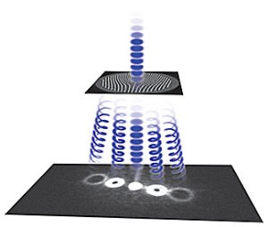
To spin up the electrons and give them orbital momentum, the NIST researchers twisted the flat electron wavefronts into a fan of helices using a very thin film with a 5-micron-diameter pattern of nanoscale slits. The pattern affects the shape of the electron wavefronts passing through it, amplifying some of the wave peaks and eliminating some of the wave valleys, to create a spiral form similar to a pasta maker extruding rotini. This method produces several electron beams fanning out in different directions, with each beam made of electrons that orbit around the direction of the beam.
The researchers knew they were successful because when they detected the electrons – which were recorded as millions of individual particles building up an image – they had formed donut-like or spiral patterns, indicating a helical shape.
Transmission electron microscopy creates images by shooting trillions of electrons through an object and measuring their absorption, deflection and energy loss. TEMs equipped with corkscrew electron beams could also monitor how the particles exert torque on a material and how a material affects the spiral shape of transmitted electrons, helping scientists build a more complete picture of the material's structure.
For example, these special electron beams have the potential to help obtain more information from magnetic materials.
"Magnetism, at its most fundamental, results from charges spinning and orbiting," McMorran said. "So an electron beam that itself carries angular momentum makes a good tool for probing magnetic materials."
A beam of corkscrew-shaped electrons, when interacting with a specimen, can exert torque on the material, by exchanging angular momentum with its atoms. In this way, the corkscrew electrons could obtain more information in the process than beams with ordinary electrons, which do not carry this orbital angular momentum.
This technique could also help improve TEM images of transparent objects like biological specimens. Biological material can be difficult to image in ordinary TEMs because electrons pass through it without deflecting. But by using corkscrew electron beams, researchers hope to provide high-contrast, high-resolution images of biological samples by looking at how the spiral wavefronts get distorted as they pass through such transparent objects.
While these imaging applications have not yet been demonstrated, producing corkscrew electrons with nanogratings in a TEM provides a significant step toward expanding the capabilities of existing microscopes.
Source: http://www.nist.gov/