The W. M. Keck Foundation has awarded Arizona State University a $1 million grant to the team of scientists led by Deirdre Meldrum at the Biodesign Institute.
The team is working to build a next-generation, 3D imaging microscope, called a “Cell-CT” scanner, that will perform functional computed tomographic (CT) imaging of individual living cells.
 Cell-CT technology will allow researchers to observe and assess the cellular function and disease status of living cells, enabling scientists to gain new insights into the metabolic pathways of disease, such as cancer.
This groundbreaking technology will allow researchers to observe and assess the cellular function and disease status of living cells, enabling scientists to gain new insights into the metabolic pathways of disease, such as cancer.
This next-generation Cell-CT scanner offers a transformative view of the biological structural and functional inter-relationships at the single cell level. Leveraging leading-edge technology developed by VisionGate, Inc. in collaboration with Meldrum’s team, this project will advance applications in basic and clinical science, deepening scientific understanding of metabolism and disease processes, and expanding the horizons of medical diagnostics.
The Biodesign Institute at ASU is boldly pushing the frontiers of science and medicine to uncover transformative solutions to the most urgent and complex challenges in human health, national security, and the well-being of our planet. Driven by passion and collaborative synergy, scientific inquiry and technology research and development, and fusing biosciences, engineering and advanced computing to fuel the translation of research advances into real solutions— Biodesign and Meldrum’s Center for Biosignature Discovery Automation (CBDA) are finding the clues that will enable us to diagnose and treat cancer sooner, in more targeted ways.
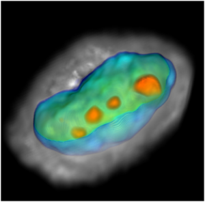
In Computed Tomography (CT), multiple 2D images taken from many perspectives are analyzed computationally to create a 3D rendering of a living structure or tissue. In diagnostic radiology, x-ray CT takes hundreds of perspective views around a single axis to create a cross sectional view through the patient’s anatomy. Similarly, the Cell-CT combines hundreds of submicron-resolution optical images taken while rotating a single cell to render a functional 3D image of the cell. That image reveals important metabolic and disease processes in action.
One of the biggest challenges in the Cell-CT scanner’s development is finding the best way to rotate cells precisely without harming them. Initially, two methods will be explored: the first rotates cells in a microfluidic vortex, and the second rotates cells with an infrared light beam. The technology will be validated through comparison studies between Cell-CT scanned cells grown in culture that represent various stages of cancers, and cells taken from human biopsies spanning the same disease spectrum.
The four-year effort is led by Professor Meldrum, who is head of CBDA at the Biodesign Institute and a leader in microdevices for biological research. Also at CBDA are Roger Johnson, an expert in instrumentation and algorithms for 3D tomographic reconstruction, who will oversee project operations and lead algorithmic and image processing research. Laimonas Kelbauskas, a specialist in laser physics and complex optical system design, will lead the optical physics and engineering team to design and build the new-generation Cell-CT system for living cells. Joining Meldrum from Turku University in Finland is Lea Sistonen, renowned expert in the effects of cell stress on signaling and gene expression. The strength of this project team is its combination of expertise and experience.
“We’re tremendously excited by the potential this technology presents for important breakthroughs, not only in cellular biology but also in medicine and ultimately personalized health care,” Meldrum said.
The Cell-CT scanner may enable, for the first time, rapid 3D spatial localization of proteins, and assessment of their concentrations in subcellular compartments and microdomains, providing powerful insights concerning relationships between cell structure and function in disease.
Source: http://www.asu.edu/