Asylum Research, the technology leader in Scanning Probe and Atomic Force Microscopy (SPM/AFM), has announced the new Electrochemical Strain Microscopy (ESM) imaging technique for its Cypher™ and MFP-3D™ AFMs.
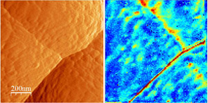 The topographic image (left) of amorphous Si anode in the Si/LiPON/LiCoO2 thin-film battery structure shows the presence of a number of grain boundaries, as well as extensive surface roughness. The ESM image (right) is obtained by measuring the electrochemical strain hysteresis loops at each pixel (100x100 pixel image over 1 micron area). The area hysteresis loop is a measure of Li-ion mobility, and is plotted as 2D map (dark blue corresponds to closed loops, red to open loops). The enhanced Li-ion mobility along the sharp grain boundary is clearly seen, as well as localized hot spots on the diffuse grain boundary and within the grains. The effective resolution of ESM for this material is ~ 10 nm, providing a high-resolution view of Li-ion dynamics in these materials. (Reprinted from N. Balke, et al., Nano Lett. 10, 3420 (2010).
Developed by Oak Ridge National Laboratory (ORNL) and Asylum Research, ESM is an innovative scanning probe microscopy (SPM) technique capable of probing electrochemical reactivity and ionic flows in solids on the sub-ten-nanometer level. ESM is the first technique that measures ionic currents directly, providing a new tool for mapping electrochemical phenomena on the nanoscale. The capability to probe electrochemical processes and ionic transport in solids is invaluable for a broad range of applications for energy generation and storage ranging from batteries to fuel cells. ESM has the potential to aid in these advances with two major improvements over other conventional technologies: (a) the resolution to probe nanometer-scale volumes and (b) the inherent ability to decouple ionic from electronic currents with (c) imaging capability extended to a broad range of spectroscopy techniques reminiscent of conventional electrochemical tools. Nina Balke of ORNL will be presenting recent results at the International Workshop on Scanning Probe Microscopy for Energy Applications (http://www.mpip-mainz.mpg.de/symposium/spm2011/) in Mainz, Germany, June 8-10 2011.
Commented Roger Proksch, President of Asylum Research, “Progress in energy storage and conversion will be greatly facilitated by the ability to study batteries and fuel cells at the level of several nanometers. ESM provides functional imaging of electrochemical phenomena in volumes millions to a billion times smaller than conventional current-based electrochemical techniques. This new technique opens the pathway to understanding energy technology and ionic devices on the level of individual grains and defects, thus bridging macroscopic functionalities and atomistic mechanisms. This in turn will lead to improved energy storage solutions – batteries with extremely high energy densities and long lifetimes and fuel cells with very high energy densities and efficiencies.”
"Traditionally, scanning probe microscopy techniques allowed measurement of electronic currents and short- and long-range forces," added Sergei Kalinin, Senior Research Staff Member in the Center for Nanophase Materials Sciences at ORNL and co-inventor (with Nina Balke and Stephen Jesse) of ESM. "ESM extends this capability to measure ionic currents, and has already been demonstrated for a variety of Li-ion cathode, anode, and electrolyte materials, as well as oxygen electrolytes and mixed electronic-ionic conductors. The ubiquitous presence of concentration-molar volume coupling in electrochemical systems suggests that this technique is in fact universal for solid state ionic imaging – from batteries and solid state to memristive electronics.
Stephen Jesse added “Perhaps even more importantly, the use of band excitation and DART engines allows measurements to be performed on rough surfaces of realistic electrochemical materials, making ESM useful for real materials and devices.”