Dec 18 2012
Scientists trying to decipher the microenvironment of living biological tissues now have a way of taking high-resolution, high-speed, three-dimensional images of their inner workings.
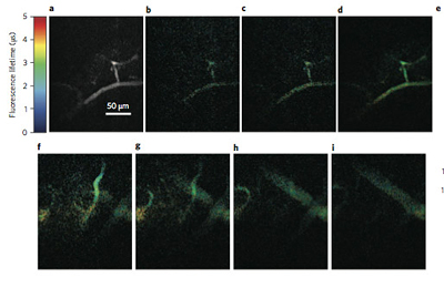 A mouse brain blood vessel is shown at different points of its phosphorescence lifetime. (Xu lab)
Cornell researchers led by Chris Xu, associate professor of applied and engineering physics, have demonstrated a new imaging technique that can quickly provide information not only about light intensity but also its "lifetime" -- how long it takes for a photon to be re-emitted after excitation.
Their technique, detailed online Dec. 16 in the journal Nature Photonics, is called frequency-multiplexed multiphoton phosphorescence lifetime microscopy, and could prove especially useful for research in biological and medical imaging.
In their experiments, they treated a mouse brain with a dye that exhibits varying phosphorescence lifetimes correlated with the amount of oxygen present in the microenvironment. This is a useful measure for biologists who, for example, look for disease states in the brain indicated by oxygen levels.
They shined a beam consisting of hundreds of discrete points, simultaneously illuminated but each encoded with a unique frequency. It is the same basic technology used in cable television, said Xu, who previously worked in telecommunications, in which each channel comes though the same coaxial cable bearing its own frequency.
"Each point in space is like an independent TV channel broadcasting the property of its own local environment," Xu said.
When each point emitted photons back at its own frequency, the researchers could then decode the lifetime of each point separately. They call this technique frequency multiplexing, and it allows them to form images by scanning hundreds of points simultaneously, rather than one at a time in existing lifetime measurement of phosphorescent dyes used in oxygen sensing.
They combined their phosphorescence lifetime mapping with multiphoton microscopy technology (invented at Cornell in 1990), which uses fluorescence to peer deep into the living tissue and build 3-D images.
Phosphorescence lifetime microscopy is intrinsically limited by the relatively slow lifetime of emitted photons in oxygen-sensitive dyes -- many microseconds, which allows researchers to only take snapshots at discrete points in the brain, rather than 3-D volume imaging.
But by simultaneously hitting the sample with hundreds of illumination points, the speed of phosphorescence-based in vivo imaging can be improved by a factor of 100 to 1,000. A process that would have taken days now takes minutes.
The work was supported by the National Science Foundation and the National Institutes of Health.
Source: http://www.cornell.edu