Jul 24 2013
Scientists from The University of Manchester have revealed new images which provide the clearest picture yet of how white blood immune cells attack viral infections and tumours.
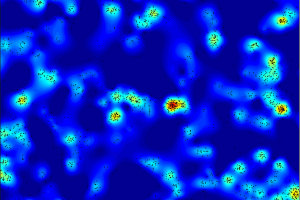 Image demonstrates how clustered these proteins are - clearly showing a nano-scale structure to how individual proteins are arranged at the surface of the Natural Killer cell. This is the first time that this level of imaging has been done for Natural Killer cells.
They show how the cells, which are responsible for fighting infections and cancer in the human body, change the organisation of their surface molecules, when activated by a type of protein found on viral-infected or tumour cells.
Professor Daniel Davis, who has been leading the investigation into the immune cells, known as natural killers, said the work could provide important clues for tackling disease.
The research, funded by the Medical Research Council (MRC) and Biotechnology and Biological Sciences Research Council (BBSRC), reveals the proteins at the surface of immune cells are not evenly spaced but grouped in clusters - a bit like stars bunched together in galaxies.
Professor Davis, Director of Research at the Manchester Collaborative Centre for Inflammation Research (MCCIR), a partnership between the University and two pharmaceutical companies GlaxoSmithKline and Astra Zeneca, said: “This is the first time scientists have looked at how these immune cells work at such a high resolution. The surprising thing was that these new pictures revealed that immune cell surfaces alter at this scale – the nano scale – which could perhaps change their ability to be activated in a subsequent encounter with a diseased cell.
“We have shown that immune cell proteins are not evenly distributed as once thought, but instead they are grouped in very small clumps – a bit like if you were an astronomer looking at clusters of stars in the Universe and you would notice that they were grouped in clusters.
“We studied how these clusters or proteins change when the immune cells are switched on – to kill diseased cells. Looking at our cells in this much detail gives us a greater understanding about how the immune system works and could provide useful clues for developing drugs to target disease in the future.”
Until now the limitations of light microscopy have prevented a clear understanding of how immune cells detect other cells as being diseased or healthy.
The team used high quality, super-resolution fluorescence microscopy to view the cells in blood samples in their laboratory to create the still images published in the journal Science Signalling this week.
Source: http://www.manchester.ac.uk/