Sep 11 2014
Comparison of 3D TEM imaging techniques reveals never-seen-before details of plant cell walls, according to a study published September 10, 2014 in the open-access journal PLOS ONE by Purbasha Sarkar from University of California, Berkeley and colleagues.
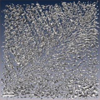 This image shows segmented cell wall volumes. Credit: Sarkar et al.
Cost-effective production of plant material for biofuel requires efficient breakdown of plant cell wall tissue to retrieve the complex sugars in the cell wall required for fermentation and production of biofuels. In-depth knowledge of plant cell wall composition is therefore essential for improving the fuel production process. The precise spatial three-dimensional organization of certain plant structures, including cellulose, hemicellulose, pectin, and lignin, within plant cell walls remains unclear, due to the limited to 2D, topographic or low-resolution imaging currently used by researchers, as well as other factors. In an attempt to compare the quality of 3D TEM imaging techniques of the cell wall structure in plant stem tissue, the authors of this study compared three different sample preparation methods for imaging: conventional microwave-assisted chemical fixation and embedding followed by imaging at room temperature; high-pressure freezing, freeze substitution (HPF-FS) followed by room temperature embedding and imaging; and cryo-immobilization of fresh tissue by self-pressurized rapid freezing, cryo-sectioning, and cryo-tomography- a type of electron microscopy run at very low temperatures that yields near-native 3D reconstructions.
Qualitative and quantitative analyses showed that plant ultrastructure and wall organization of cryo-immobilized samples were preserved remarkably better than conventionally prepared samples. However, due to the highly challenging techniques associated with cryo-tomography, large-scale quantitative analyses are better performed on HPF-FS samples.
Manfred Auer added: "We have developed and compared novel sample preparation and molecular 3D imaging approaches for plant cell walls, yielding insight into faithfully preserved 3D wall architecture, which will lead to rational re-engineering of second-generation lignocellulosic biofuel crops."
Source: http://www.plos.org/