May 26 2015
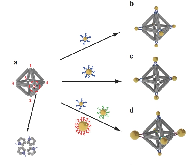 Scientists built octahedrons using ropelike structures made of bundles of DNA double-helix molecules to form the frames (a). Single strands of DNA attached at the vertices (numbered in red) can be used to attach nanoparticles coated with complementary strands. This approach can yield a variety of structures, including ones with the same type of particle at each vertex (b), arrangements with particles placed only on certain vertices (c), and structures with different particles placed strategically on different vertices (d).
Researchers at Brookhaven National Laboratory of the U.S. Department of Energy (DOE) have combined the artificial strands of DNA to function in two ways: rope-like configurations of the DNA double helix were used to create a strong geometrical framework, and dangling pieces of single-strands of DNA were added to stick nanoparticles in place.
The study has been described in the journal Nature Nanotechnology. This is a new breakthrough on the use of DNA in nanoscale construction. The method resulted in arrays and clusters of nanoparticles, which represent a major milestone for designing materials with customized functions and structures for applications in medicine, optics, and energy.
These arrays of nanoparticles with predictable geometric configurations are somewhat analogous to molecules made of atoms. While atoms form molecules based on the nature of their chemical bonds, there has been no easy way to impose such a specific spatial binding scheme on nanoparticles. This is exactly the problem that our method addresses, said Brookhaven physicist Oleg Gang, who headed the project at the Lab's Center for Functional Nanomaterials (CFN), a DOE Office of Science User Facility.
According to the researchers, the novel technique would allow them to organize the arrangements of a variety of nanoparticles and help them manipulate the synergistic or collective effects. Some of the examples comprise materials that deliver biomolecules, rotate light, or control the flow of energy. The researchers designed the nanoparticle architectures by means of an octahedral scaffold, with particles placed in accurate locations on the scaffold as per the specificity of DNA coding. These designs contained two varied arrangements of the same group of particles, with each configuration having different optical properties. Geometrical clusters were also utilized as building blocks for larger arrays such as2D-planar sheets and linear chains.
We may be able to design materials that mimic nature's machinery to harvest solar energy, or manipulate light for telecommunications applications, or design novel catalysts for speeding up a variety of chemical reactions," Gang said.
Our work demonstrates the versatility of this approach and opens up numerous exciting opportunities for high-yield precision assembly of tailored 3D building blocks in which multiple nanoparticles of different structures and functions can be integrated," stated Ye Tian, CFN scientist and one of the lead authors of the paper.
This method of nanoscale construction leverages two major properties of the DNA molecule, such as the natural tendency of strands having complementary bases, and the double helix, twisted-ladder shape to combine in an accurate way. Initially, bundles of six double-helix molecules were produced, with four of these bundles combined together to form a stable and a relatively strong building material, just like the way fibrous strands are integrated together to form a rigid rope. These rope-like girders were then utilized to create the frame of 3D octahedrons, eventually combining the linear chains of DNA with countless number of short complementary strands of DNA. These were known as DNA origami octahedrons.
In order to attach the nanoparticles to the 3D frames, the researchers designed each of the six-helix bundles in such a way that a single helix had an additional piece of single-stranded DNA sticking out from either ends. When each vertex of the frame was organized into 3D octahedrons, some of these sticky end tethers were available in each vertex for adhering with objects covered with complementary strands of DNA.
When nanoparticles coated with single strand tethers are mixed with the DNA origami octahedrons, the 'free' pieces of DNA find one another so the bases can pair up according to the rules of the DNA complementarity code. Thus the specifically DNA-encoded particles can find their correspondingly designed place on the octahedron vertices, said Gang.
By altering the sequences of DNA encoded on the tethers, the researchers can modify what binds to the individual vertex. In one such experiment, the same sequence was encoded on all tethers of the octahedron, and the strands were fixed with a complementary sequence to gold nanoparticles. This resulted is a single gold nanoparticle fixed to individual octahedron's six vertices.
In further experiments the sequence of certain vertices was altered and complementary strands were utilized on a range of particles. This showed that both the arrangement and assembly of the particles can be controlled in an accurate manner. In a similar experiment, two different arrangements were made from the same three pairs of particles having different sizes, which resulted in products having varied optical characteristics. The team also used DNA tethers on specified vertices to join octahedrons end to end and in 2D arrays, creating chains and sheets, respectively.
However, a major challenge was to validate the arrangement and structure of particles. This is because the DNA molecules and nanoparticles, which constitute the frames, exhibit different densities. While some microscopy methods can show the particles alone, others would change the 3D structures.
The team employed cryo-electron microscopy, also known as cryo-EM, to observe the particles and origami frames. This work was headed by Huilin Li, Brookhaven Lab and Stony Brook University biologist, and Tong Wang, the other lead co-author of the paper and who also works with Li in the Biosciences department of Brookhaven. In order to view the varied density components individually, the researchers need to subtract the data from the images and integrate the same by means of single particle 3D reconstruction and tomography to create the final images. The images thus obtained showed that the new method used to direct the placement of nanoparticles on DNA-encoded vertices of molecular frames can prove to be effective for designing new nanomaterials.
Cryo-EM preserves samples in their near-native states and provides close to nanometer resolution. We show that cryo-EM can be successfully applied to probe the 3D structure of DNA-nanoparticle clusters, Wang said.
The DOE Office of Science supported the study. DOE’s Office of Science supports the Brookhaven National Laboratory. It supports fundamental studies on the physical sciences in the US and is pursuing to tackle the most pressing complexities of today’s time.