Oct 26 2015
For a project funded by the Bill and Melinda Gates Foundation’s Grand Challenges in Global Health Initiative, a research team from Rice University has developed a plastic, miniature digital fluorescence microscope that can be used in rural areas to quantify white blood cell levels in patients.
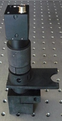 The assembled 3-D printed WBC microscope rests on an optical bench. The holes on the bench are spaced 1 inch apart, giving an idea of the microscope's size. Credit: Alessandra Forcucci, Rice University
Doctors can learn a lot about the state of a patient’s immune system just by examining their blood under the microscope. An abnormally high or low white blood count, for instance, might indicate a bone marrow pathology or AIDS. The rupturing of white blood cells might be the sign of an underlying microbial or viral infection. Strangely shaped cells often indicate cancer.
While this old, simple technique may seem a quaint throwback in the age of high-tech health care tools like genetic sequencing, flow cytometry and fluorescent tagging, the high cost and infrastructure requirements of these techniques largely limit them to laboratory settings — something point-of-care diagnostics aims to fix.
The plastic microscope developed by Rice researchers is intended for use in parts of the world far removed from the modern laboratory.
“One of the driving aspects of the project is the cost of the sample or sample preparation,” said Tomasz Tkaczyk, associate professor of bioengineering. “Many systems which work for point-of-care applications have quite expensive cartridges. The goal of this research is to make it possible for those in impoverished areas to be able to get the testing they need at a manageable price point.”
One of Tkaczyk’s co-authors on the research was Rebecca Richards-Kortum, Rice’s Malcolm Gillis University Professor, director of the Institute of Biosciences and Bioengineering and of Rice 360°: Institute for Global Health Technologies and a fellow at The Optical Society.
How the microscope works
The researchers’ device identifies and quantifies three types of white blood cells –lymphocytes, monocytes and granulocytes — in a drop of blood mixed with a staining compound. The compound is repelled by water at neutral pH, which allows it to easily diffuse through cellular and nuclear membranes, where it turns green or red when encountering DNA or RNA, respectively, with emission maximums at 525 nanometers and 650 nanometers. By optimizing a microscope for these emission peaks, the researchers are then able to quantify the white blood cells in a sample consisting only of 20 microliters of dye, 20 microliters of whole blood and a glass slide with a coverslip.
“You can just count cells,” Tkaczyk said. “The device doesn’t require the highest resolution, because we’re not really focused on morphology here, but it needs to be able to resolve single cells, which are on the order of one micron or more.”
The ratio in color allows the researchers to differentiate among monocytes, granulocytes and lymphocytes, which is significant because quantifying this three-part white blood cell count is an essential first step in diagnosing a number of disorders.
The researchers used a single-point diamond-turning lathe to fabricate the part of the microscope called the objective, which consists of one polystyrene lens and two polymethyl methacrylate aspheric lenses. The lenses were then enclosed in an all-plastic, 3-D-printed microscope housing and objective. Once constructed, the microscope provided a field of view of 1.2 millimeters, allowing for at least 130 cells to be present for statistical significance when quantifying white blood cells. Once constructed, the microscope doesn’t require any manual adjustment between samples.
The prototype microscope, which also includes an LED light source, power supply, control unit, optical system and image sensor, cost less than $3,000 to construct. At production levels upward of 10,000 units, the researchers estimate that this price would fall to around $600 for each unit, with a per-test cost of a few cents.
Future work for Tkaczyk and his colleagues includes developing an automated algorithm for identifying white blood cells, as well as comparing their differential counts to other conventional hemotology analyzers. The use of low-cost components such as LEDs, reflectors and USB detectors, combined with the all-plastic housing and lenses, will allow future versions of the prototype to be mass-produced.
Source: http://www.rice.edu/