Jan 28 2016
A team of researchers from the California NanoSystems Institute at UCLA have developed a new method, known as wavelength scanning pixel super-resolution, which improves digital microscopy images.
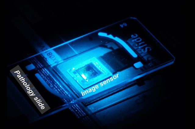 The image sensor of the wavelength scanning super-resolution apparatus collects a “stack” of images of the sample. (Credit: Ozcan Lab)
This new method is important because digital imagery has resulted in a number of developments in the field of microscopy, but digital microscopic imaging can sometimes produce pixelated images that are blurry.
Wavelength scanning pixel super-resolution utilizes a device that captures a stack of digital images containing the same specimen. Each image has a slight different wavelength of light. The research team then applied a recently developed algorithm, which divides the pixels present in each captured images into several smaller pixels. This delivers a digital image of the specimen with a higher resolution.
The research team was headed by Aydogan Ozcan, Chancellor’s Professor of Electrical Engineering and Bioengineering at the UCLA Henry Samueli School of Engineering and Applied Science. The study was reported in Light: Science and Applications, a journal from the Nature Publishing Group.
These results mean we can see and inspect large samples with finer details at the sub-micron level. We have applied this method to lens-based conventional microscopes, as well as our lensless on-chip microscopy systems that create microscopic images using holograms, and it works across all these platforms.
Aydogan Ozcan, Professor, Electrical Engineering and Bioengineering, UCLA
This new technique has a wide range of benefits, especially in the field of pathology, which deals with very fast microscopic imaging of multiple blood and tissue cells for diagnosing diseases like cancer. The study used samples of blood to screen different diseases and also conducted Papanicolaou tests, used to screen for cervical cancer.
Ozcan stated that wavelength scanning super-resolution is suitable for samples that are both dye-stained and colorless. The equipment is positioned entirely on a desktop, meaning the size and convenience of this equipment are both beneficial to scientists and doctors utilizing microscopes in places that have very limited resources, for example, clinics operating in developing countries.
The first author of the study was UCLA graduate student Wei Luo. The other authors were graduate students Yibo Zhang and Alborz Feizi and postdoctoral scholar Zoltan Gorocs.
The researchers carried out some experiments at the Nano and Pico Characterization Lab at the California NanoSystems Institute at UCLA.
The research was supported by the Presidential Early Career Award for Scientists and Engineers, the Army Research Office, the National Science Foundation, the Office of Naval Research and the Howard Hughes Medical Institute.
Source: http://www.ucla.edu/