Jun 24 2008
Life is strongly related to the behavior and interactions of individual molecules. Molecules bind together forming aggregates, compartments, cells, tissues, organs and organisms. Molecular recognition as the base mechanism of binding events happens fast and on the nm scale where Brownian motion is the driver of molecular binding processes. The concept of using nanoparticles as a local sensor or probe enables important developments in medicine, molecular and cellular biology. To observe the entrance process of a particle into a cell in real time, in 3D and without perturbation of the biological specimen is a dream of many life science researchers. Existing technologies such as epifluorescence, TIRF, LSM and video particle tracking have several drawbacks. Labelling is time consuming and causes perturbation in single molecule experiments. The best resolution in confocal microscopy (CLSM) is ~300nm in the X & Y and ~500nm in the Z axis. Fluorescence i s not label free and has poor performance for processes occurring on cell membranes. TIRF measures only the first 200nm from the surface of the cell and gives no access to the whole cell volume. Particle tracking by video microscopy is only a 2D technique with best resolution of 15nm.
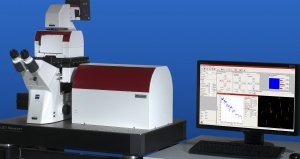 The NanoTracker™ head assembly with mounted fluid cell
What kind of tool is needed to fulfill the dream? The answer is a live cell imaging technique with high temporal and spatial resolution applied to non labelled nanoparticles. JPK Instruments’ new NanoTracker™ optical tweezers and 3D particle tracking system delivers live cell imaging at a new level. With the NanoTracker™, the user can trap and track particles from several µm down to 30nm with the ability to control, manipulate and observe vesicles, endosomes, gene and drug spheres, viruses and bacteria, nanoprobes or -carriers, biomarkers or even whole cells in real time with nanometer precision. This opens up new applications in many different disciplines including biophysics, biochemistry, cellular and medical research in microbiology, developmental- and system biology, infection research and immune response, toxicity of nanoparticles and many more.
There are many potential application areas for applying an optical tweezers platform. These include the study of single molecules & biopolymers, cell membranes, cell-particle interaction and viral and bacterial infection.
Up until this time, the majority of optical tweezers systems have been built by researchers for their own needs. There are variants consisting of different laser set-ups, single or multiple optical traps and different optical detection techniques. JPK’s NanoTracker™ offers the first system with full integration into an inverted research microscope and complete environmental control of the sample. In general, experiments require long-term system stability and this is achieved with the choice of an ultra- stable 1064nm laser, a folded optical pathway in combination with a drift-compensated design.
You are invited to visit JPK at MicroScience 2008 in London, June 24 to 26, and see the system on stand H8.