Jan 26 2018
According to researchers at Washington University School of Medicine in St. Louis, a new anti-cancer approach wields light as a precision weapon. Unlike traditional light therapy — which is restricted to the skin and areas accessible with an endoscope — this method can target and attack cancer cells that have spread deep inside the body.
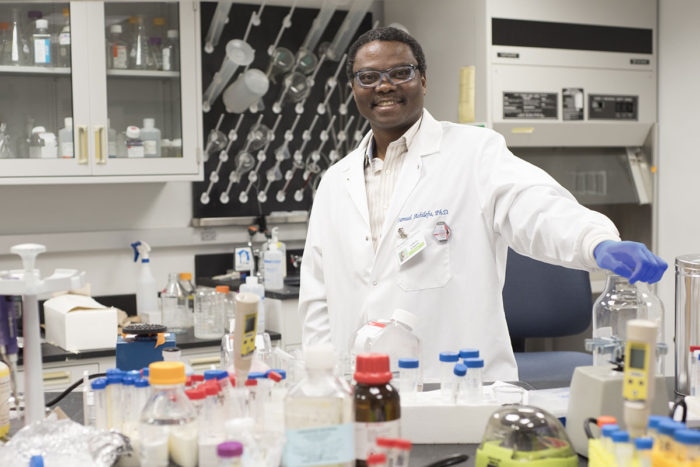 Samuel Achilefu, PhD, and his colleagues are working to develop a novel cancer therapy that uses light against tumors that have spread. (Image credit: Washington University School of Medicine)
Light released as part of traditional cancer-imaging methods to detect metastatic tumors, can also activate light-sensitive drugs according to the new research. Furthermore, the research reveals that when such drugs are loaded into nanoparticles that target lit-up cancer cells, the light-sensitive drug creates toxic free radicals that destroy the tumor cells. The team demonstrated that the method worked well in mice with multiple myeloma, a cancer of white blood cells, and aggressive metastatic breast cancer.
The research details can be found online in Nature Communications.
“Cancer that has spread remains the major reason patients die,” said senior author Samuel Achilefu, PhD, the Michel M. Ter-Pogossian Professor of Radiology at the School of Medicine. “Our study shows that this phototherapeutic technology is particularly suited to attacking small tumors that spread to different parts of the body, including deep in the bone marrow.”
The technology harnesses a chemotherapy drug known as titanocene. As a chemotherapy agent alone, titanocene has not functioned well in clinical trials, even at comparatively high doses. But when exposed to the radiation discharged by visible light, titanocene creates reactive particles that are toxic to cells, even at low doses.
Achilefu and his colleagues packaged low doses of titanocene into nanoparticles they targeted to proteins believed to sit on the surface of cancer cells. They discovered that when the nanoparticles make contact with cancer cells, their membranes fuse together, discharging the titanocene into the cells.
The researchers then deliver a common cancer imaging agent known as fluorodeoxyglucose (FDG), a type of sugar. Energy-hungry cancer cells take up the FDG at high rates, causing tumors to glow in a positron emission tomography (PET) scan. This glow also activates the titanocene, discharging free radicals and destroying the cells.
Since the titanocene and the light-emitting FDG are targeted to the same place at the same time exclusively in tumors, the method is thought to be less toxic than chemotherapy and standard radiation. Research also reveals that the body rids itself of titanocene through the liver, while FDG is sent out via the kidneys. As the two components are disposed of separately, it reduces damage to other organs. When kept apart, the two components are not lethal, according to the researchers.
Once a week for four weeks, mice with multiple myeloma were treated using this approach. In the weeks following, the treated mice had considerably smaller tumors and survived longer than the control mice. Fifty percent of treated mice survived a maximum of 90 days. Of the control mice, 50 % survived 62 days. The mice with breast cancer also exhibited an anti-tumor effect when treated using this approach, though less pronounced than in those with multiple myeloma, possibly because of the extreme aggressiveness of the breast cancer cell line, according to the researchers. The team also discovered that some types of multiple myeloma unexpectedly were resistant to this procedure. They established that the resistant multiple myeloma cells lacked the surface proteins used to target the titanocene-loaded nanoparticles.
This is an opportunity to learn because it’s similar to what is seen in patients — some of the cells become dormant but don’t die after treatment. When we looked closer at the cells that were resistant to our phototherapy, we saw that the surface protein we are targeting was not there. So next, we want to find out if we can pinpoint another surface protein to target and kill these resistant cells along with the myeloma cells that did respond to the original therapy, which could lead to complete remission.
Samuel Achilefu, Senior Author
Achilefu foresees doctors being able to use this type of technology in the future to prevent cancer from recurring.
We are interested in exploring whether this is something a patient in remission could take once a year for prevention. The toxicity appears to be low, so we imagine an outpatient procedure that could involve zapping any cancerous cells, making cancer a chronic condition that could be controlled long-term.
Samuel Achilefu, Senior Author