A UV/Visible spectrophotometer equipped with a 150 mm integrating sphere and center mount can make numerous measurements significant to the nanomaterial industry.
This article covers a technique to differentiate between large particle scattering, nanoparticle scattering, and sample absorbance. Moreover, the nanoparticle characterization technique enables measurements on finished products, such as liquids, films, solid gels, powders and creams.
It is also useful in formulation studies, QC analysis of finished products, and individual product component analysis, and shows promise for use in a myriad of marketing segments across the nanomaterials industry with possibility in food, environmental, industrial and medical/pharmaceutical applications.
The technique needs to include the characterization and quantitation of nanoparticle scatter (< 700 nm), large particle scatter (>700 nm), and absorbance. It is possible to characterize the samples utilizing the UV/Visible spectrophotometer fitted with a 150 mm standard integrating sphere and center mount. Figure 1 depicts the optical diagram for both the reflection and transmission measurements of the double beam integrating sphere.
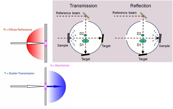
Figure 1.
The transmitted and forward-scattered light is collected by placing the sample in front of the sphere at the scatter transmission port, as shown in Figure 1 on the left in blue.
By placing the sample on the opposite side of the sphere at the diffuse reflectance port, the diffusely reflected and back-scattered light is collected, as illustrated in Figure 1 on the left in red. All of the scattered light from the sample is received and measured between these different measurements.
However, there is a possibility for light to be lost due to absorption by the sample in these scatter transmission and diffuse reflectance measurements, as shown in the pink colored regions of the sample in Figure 1 on the left.
Instrumentation
A PerkinElmer Lambda 1050 UV/Vis/NIR instrument equipped with the 150 mm sphere and center into its detector compartment area is illustrated in Figure 2. The scatter transmission port positioned on the left hand side of the sphere can be seen in the center of the picture.
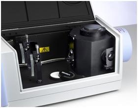
Figure 2.
Part of the reference and sample beam transfer optics can be spotted on the left. The diffuse reflectance port is under the blue cover and on the right of the sphere. The sphere is equipped with a center mount accessory which can be spotted on top and in the center of the sphere housing in Figure 2.
The key component of this analysis is the center mount, which is shown in Figure 3. A “clip type” center mount shown in the top diagram of Figure 3 is designed for holding many different flat samples. The “paddle” assembly at the bottom of the sphere prevents the entry of the light scattered from the sample into the detectors.
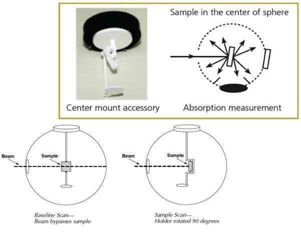
Figure 3.
The light from the sample needs to be reflected from the interior of the sphere for multiple times. The top right diagram in Figure 3 depicts the operation of the center mount/sphere configuration to receive all of the sample-scattered light from all angles.
A center mount sampling background, or a baseline measurement is taken with a sample in the center mount, which is turned for preventing the illumination of the beam, as shown in the bottom left of Figure 3.
The bottom right diagram of Figure 3 depicts the sample measurement setup with the sample oriented in such a way that the instrument beam makes an 8° angle of incidence with the sample.
Analysis
The transmission, reflection, and center mount spectra for a Transpore Surgical Tape sample is shown in Figure 4. It is exemplary of typical data for this analysis. This configuration can corroborate that the center mount does indeed receive collect all of the scattered light. This can be done by adding together the scatter transmission and diffuse reflectance spectra, which are then compared with the center mount spectrum.
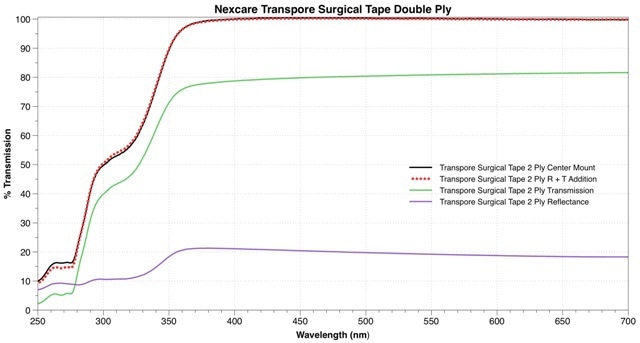
Figure 4.
After completely collecting both the forward and backscattered light in the scatter transmission (solid green line) and diffuse reflectance (solid purple line) spectra respectively, it is necessary to add them up to the center mount spectrum.
The graph shown in Figure 4 illustrates spectra from a two ply layer of surgical tape, which is a synthetic substrate utilized to mimic the spectral properties of human skin. The addition product of the reflectance and transmission spectra is represented as the red dotted line, which overlays and compares with the center mount spectrum in solid black.

This information has been sourced, reviewed and adapted from materials provided by PerkinElmer Inc.
This article was adapted with kind permission from work provided by Jeffrey L. Taylor and Jillian F. Dlugos from PerkinElmer.
For more information on this source, please visit PerkinElmer Inc.