WITec’s alpha300 R confocal Raman microscope series has long been recognized as the benchmark in advanced Raman imaging systems. Continuous development driven by WITec’s innovative spirit keeps its range of capabilites at the industry's leading edge.
The alpha300 R offers unparalleled speed, sensitivity, and resolution. These qualities are available simultaneously and without compromise, which allows the quick investigation of even weak Raman scatterers and extremely low material concentrations or volumes with the the lowest excitation energy. Optimized optical components enable Raman spectra to be recorded at each image pixel with acquisition times on the order of milliseconds.
Due to the confocal setup, it is not only possible to collect information from the sample's surface, but also to look deep inside transparent samples and obtain 3D information in depth scans and image stacks. When analyzing dedicated peak characteristics of the spectra, a variety of images can be generated using only a single set of data. This allows the distribution of chemical compounds, crystallinity, or material stress properties to be visualized. In addition to the imaging capabilities, the system can also be used to collect Raman spectra at selected sample areas and along arbitrary lines or to acquire time series.
The alpha300 R series' inherent modularity makes it possible to combine Raman imaging with complementary imaging methods including AFM, SNOM and SEM, and allows it to evolve with changing experimental requirements. Applications include materials science, coating and thin film analysis, geoscience, pharmaceutics, food science and many others.
Key Features of the alpha300 R
- Confocal Raman imaging with unmatched performance in sensitivity, speed, and resolution
- Industry-leading lateral resolution
- Hyperspectral image generation through the acquisition of a complete Raman spectrum at every image pixel
- Ultra-fast Raman imaging available with below 1 ms integration time per spectrum
- Non-destructive imaging method: Staining or specialized sample preparation unnecessary
- Ultra-high throughput spectroscopic system for maximum sensitivity and outstanding spectral resolution
- Exceptional depth resolution ideally suited to 3D image generation and depth profiles
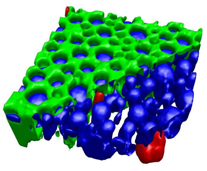
Confocal 3D Raman volume image of a pharmaceutical emulsion: The oil phase (green) is partially removed in the image for better visibility of the silicon impurities (red) in the water and API containing phase (blue). Image Credit: WITec GmbH
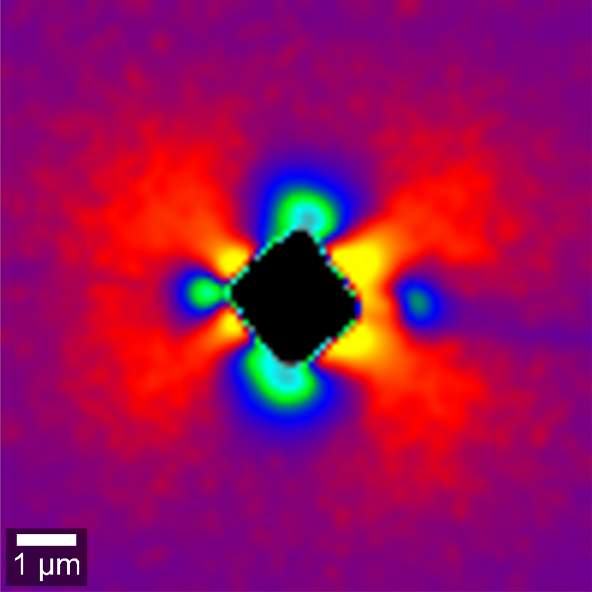
Material stress in silicon imaged via Raman peak-shift analysis. Tensile strain (blue) and compressive strain (yellow). Image Credit: WITec GmbH
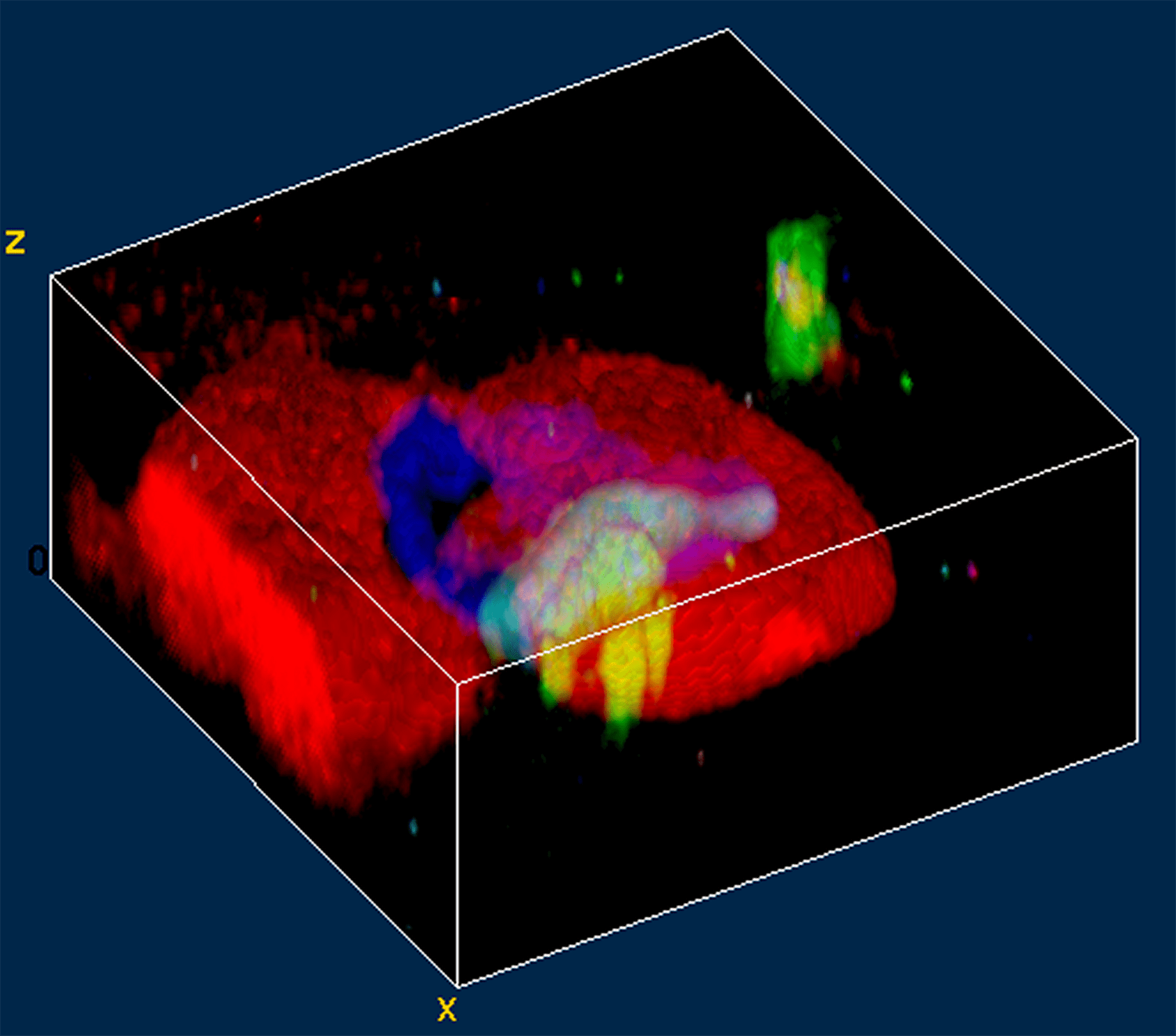
3D Raman image of a fluid inclusion in garnet. Image Credit: WITec GmbH