Dec 3 2009
Olympus, the world leader in objective-based total internal reflection fluorescence (TIRF) microscopy, has announced another significant leap forward in multicolor TIRF imaging. The Olympus cell^TIRF™ illuminator offers four motorized channels for simultaneous image capture; intuitive, user-friendly software control of TIRF parameters; and instant setting and confirmation of the precise TIRF angle. The system also allows easy transition back and forth to widefield fluorescence and a sleek, space-saving ergonomic design.
The new system has four individually controlled motorized laser inputs for TIRF imaging. With the cell^TIRF system, each laser wavelength is optimally focused and each angle is individually set, allowing different wavelengths to have the same penetration depth. Combined, these features make cell^TIRF the only such system that can simultaneously capture multiple channels with independently adjusted TIRF angles. Researchers do not have to make time-consuming adjustments during experiments.
Users can operate the software via a simple graphical user interface (GUI), keyboard arrow keys or the mouse wheel, making it easier than ever to control the incident angle of each wavelength and adjust TIRF penetration depths. Best of all, users can preset calculated penetration depths for all four lasers with just a single mouse click; the system will individually adjust each laser’s angle automatically to simultaneously capture TIRF from all four channels.
A number of additional capabilities make the new system especially useful for cell biologists and other researchers. For instance, one laser line can be adjusted so the system can do fluorescence recovery after photobleaching (FRAP) experiments. A handy button is available to seamlessly switch to widefield imaging, allowing researchers to visualize the complete cell profile, see nuclei or find the field they want to observe. TIRF imaging mode can be reestablished in less than 500msec. Users can also control laser intensities easily onscreen.
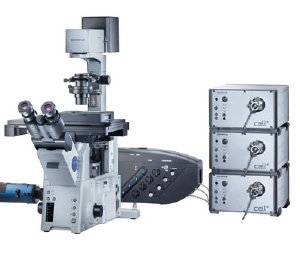
The sleek, elegant design has all four laser inputs coming in from one side, making the compact system easy to set up and integrate with incubator systems. Researchers can quickly insert clean-up filters for lasers if needed. In addition, the main unit is built from single billet aluminum for rigidity and robustness, making it more tolerant of small temperature fluctuations that may occur in rooms where it is placed.
Accompanying the launch of the cell^TIRF illuminator are new Olympus laser systems incorporating all the most commonly used laser wavelengths in the 405 – 640nm range. The directly coupled laser systems feature attenuable laser power up to 100mW, and offer better delivery efficiency to the microscope than laser combiner options. Each laser system is a small, stackable integrated unit.
“Olympus was the first company to commercialize TIRF microscopy, and as the pioneer in this field, we’ve taken another key step with advanced motorization, additional inputs for simultaneous TIRF acquisition, and a great GUI to easily and repeatably set incident angles,” said Stuart Shand, TIRF marketing manager for Olympus America Inc.
The introduction of the cell^TIRF system builds on Olympus’ market-leading tradition in TIRF. Olympus introduced the first TIRF microscope objective in the U.S. in 1997, and continues to offer the largest line of TIRF optics, including the unmatched APO 100x NA 1.65 objective that can achieve penetration depths below 50nm. Other TIRF objectives include the APO N 60x NA 1.49, PlanApo 100x NA 1.45, and U-APO 150x NA 1.45. The new system can be retrofitted to Olympus IX2® microscopes including the IX71 and IX81 microscopes.
TIRF imaging was developed to deliver information about the surfaces of cells where vital functions such as communication, absorption, signaling, growth and movement occur. TIRF imaging occurs where the cell membrane comes in contact with a cover slip. The laser illumination light’s angle of incidence dictates how deeply an evanescent wave penetrates into the cell, and lasers of different wavelengths require varying angles to operate consistently with each other. Olympus’ ability to do simultaneous multichannel image capture allows scientists to efficiently study rapidly changing intracellular processes, do high-precision ratio experiments, and more.