An innovation that can help scientists observe a reaction moving at greater than 10 meters per second, with a few nanometers spatial resolution, is a feat some would say is nearly impossible.
But not the Lawrence Livermore team of scientists who developed the dynamic transmission electron microscope (DTEM).
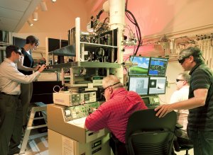 Working with the dynamic transmission electron microscope (DTEM). From left: Bryan Reed, Melissa Santala, William DeHope, Thomas LaGrange, Joseph McKeown. Photo by Jacqueline McBride/LLNL
DTEM’s ability to let researchers peer into the heart of scientific phenomena while it’s happening has earned it one of the 10 winning microscopy innovations in the 2010 Microscopy Today Innovation Award competition.
Microscopy Today's MT-10 Awards recognize the best new products and methods across the entire field of microscopy. Five of the awards are primarily related to the life sciences and five are related to the physical sciences. In each of these areas, there may be interesting new developments in light microscopy, scanning probe microscopy, electron microscopy, ion microscopy, acoustic microscopy, microanalysis, specimen preparation, etc. These awards honor the best developments in microscopy from the previous calendar year.
The award will be given to the team at the 2010 Microscopy & Microanalysis meeting held Aug. 1-5 in Portland, Ore. Descriptions of the winning products and methods will be published in the print and digital editions of the September 2010 issue of Microscopy Today.
Unlike traditional transmission electron microscopes that are generally restricted to capturing images before and after some rapid transformation (such as a material deforming or the growth of a nanowire), the DTEM captures images during the process itself. DTEM goes beyond merely revealing that a transformation has happened; it provides crucial details of how, when and where it happened. For example, while a conventional electron microscope can produce static images of viruses before and after they have attacked cells, the DTEM could potentially capture a virus in the process of joining to a membrane and releasing its genetic material in a rapid sequence of short-exposure images.
The DTEM is able to take snapshots of the dynamics that occur in samples of material under strenuous conditions – extreme temperature, applied pressure, surface corrosion – creating a visual record of microstructural features as they rapidly evolve.
It combines all of the powerful techniques of the standard TEM with nanosecond time resolution for capturing dynamic processes while they occur with single-shot measurements. (The term “single shot” means the gathering of the required data, diffraction pattern or image, using only one bunch of electrons.)
The Livermore microscope already has produced new levels of scientific understanding of nanostructure growth, phase transformations and chemical reactions. But this is only the beginning.
DTEM provides an entirely new way of exploring material processes with a range of potential applications that have just been undertaken.
In a recent experiment, the team was able to peer into the inner workings of catalyst nanoparticles 3,000 times smaller than a human hair within nanoseconds.
The findings point the way toward future work that could greatly improve catalyst efficiency in a variety of processes that are crucial to the world’s energy security, such as petroleum catalysis and catalyst-based nanomaterial growth for next-generation rechargeable batteries.
The research is funded by the Department of Energy’s Office of Science, Office of Basic Energy Sciences, Division of Materials Sciences and Engineering.
Members of the team include: Wayne King, Michael Armstrong, Nigel Browning, Geoffrey Campbell, William DeHope, Judy Kim, Thomas LaGrange, Benjamin Pyke, Bryan Reed, Richard Shuttlesworth, Brent Stuart and former LLNL employees J. Bradley Pesavento Mitra Taheri and Benjamin Torralva.