Jun 19 2012
NanoSight, leading manufacturers of unique nanoparticle characterization technology, reports on the work of Professor Hang (Hubert) Yin's group at the University of Colorado at Boulder where they apply Nanoparticle Tracking Analysis (NTA) to characterize biological nanoparticles such as microvesicles.
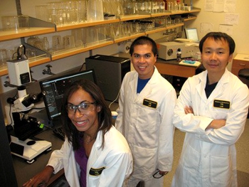 Professor Hang "Hubert" Yin with two members of his research group, Leslie-Anne Morton and Jonel Saludes, use the NanoSight LM10 system.
The Yin Research Lab is interested in studying at the interface of chemistry, Biology and engineering with a particular focus on structure-based drug design, cell signaling, biochemistry, biotechnology development and membrane protein simulations.
The main research goal of the group is to identify and design peptides that sense membrane curvature to better understand protein/peptide-lipid interactions and potentially create non-invasive probes to detect highly curved extracellular vesicles. Currently, we are studying microvesicles as potential biomarkers of tumor progression and cancer metastasis. These nanoparticles are shed into bodily fluids targeting other cells in the body and are vital for inter-cellular communication.
Their experimental protocol involves lipid vesicle preparation by pressure-controlled extrusion through different membrane pore sizes. Different lipid vesicle sizes are prepared in order to mimic the size range of the microvesicles that are shed into the extracellular matrix. Following vesicle extrusion, it is important to validate the vesicle size. By using Nanoparticle Tracking Analysis (NTA) technology, the results provide an accurate quantification of different populations of vesicle sizes present in the sample.
Prior to NTA, the group mostly used dynamic light scattering (DLS) to determine the sizes of our synthetic lipid vesicles. Speaking on their use of NTA, Professor Yin says "NTA brought several benefits over existing methods. The detection ranges from 10 - 2000 nm for vesicle sizes, dimensions that cover our liposome size of interest. Flow cytometry has a lower limit detection of ~200 nm to accurately measure particle sizes so did not reach our lower requirement while DLS measures the average size of all the particles present in the sample rather than accurately distinguish different pools of vesicle sizes, often creating a bias towards larger particles."
The group has recently published a paper in the Journal of Visualized Experimentation that used the NTA technology entitledConstant Pressure-controlled Extrusion Method for the Preparation of Nano-sized Liposomes (Leslie A. Morton, Jonel P. Saludes, Hang Yin). To learn more, please refer to doi: 10.3791/4151.