Sponsored by MerckReviewed by Emily MageeFeb 1 2024
Iron oxide-incorporated conjugated polymer nanoparticles, also known as conjugated polymer nanoparticles (CPNs), are highly fluorescent particles composed of a semiconductor light-emitting polymer core enveloped by a biocompatible surfactant.
These new bioimaging probes possess remarkably brighter fluorescent properties compared to other markers. Their intense brightness stems from exceptional extinction coefficients and superior stability against light, heat, and chemicals.1
CPNs also exhibit no toxicity, making them compatible with live cell systems.2,3 The CPN core also contains iron oxide, allowing for magnetic manipulation, making them excellent agents for multi-modal MRI imaging. Refer to Table 1 for a comprehensive list of CPN attributes.
Table 1. List of CPN properties. Source: Merck
| Property |
Value |
| Size |
80 nm |
| Thermal stability range |
-80 °C – 120 °C |
Photostability duration
(ambient temperature and illumination) |
24 months |
| Photostability duration (laser excitation) |
>6 hours |
| pH stability range |
pH 2.0-12.0 |
| Magnetic |
Overnight pull down |
| Linkage chemistry |
EDC (-COOH on CPN and –NH2 on target) |
| Zeta potential |
-40 mV |
| Suspension buffer |
Water |
CPNs cover fluorescence emission wavelengths ranging from 420 nm to 680 nm in the visible spectrum. This wide spectral range enables CPNs to be used in various applications such as flow cytometry, immunohistochemistry, ELISA, and lateral flow devices, aligning with standard filter sets and laser lines.
These functionalities are facilitated by surface-accessible carboxyl groups, enabling the binding of diverse molecules like antibodies, proteins, streptavidin, and nucleic acids (Figure 1).

Figure 1. Linkage scheme: CPNs linked via carboxyl groups to amino groups on targeting molecules, such as antibodies. This reaction is mediated by the use of 1-ethyl-3-(3-dimethylaminopropyl)carbodiimide (EDC). The ratio used in the standard linkage protocol would attach approximately 40 antibody IgG to each CPN. Image Credit: Merck
Characteristics of CPNs
Structural Properties
The inclusion of LEP within the shell of a biocompatible surfactant creates a highly hydrophilic structure, forming water-soluble micelles of various sizes (Figure 2).
This 'core-shell' arrangement (Figure 3) offers an ideal surface for attaching functional molecules like streptavidin, antibodies, targeting proteins, or nucleic acids through different surface chemistry methods.
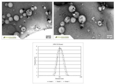
Figure 2. CPNs display excellent uniformity in size and shape, as shown in the Transmission Electron Microscopy (TEM) above, and Dynamic Light Scattering (DLS) below. Image Credit: Merck
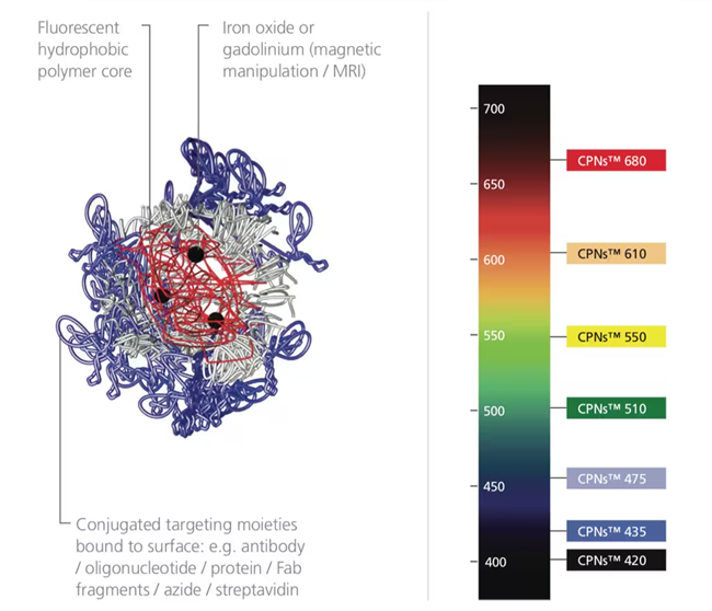
Figure 3. CPN structure alongside a light spectrum labeled with the range of CPNs available. CPNs absorb with a notable Stokes shift, larger than many organic dyes. Image Credit: Merck
Integrating iron oxide into the core enables the manipulation of attached molecules or cells using magnetism (Figure 4), aiding in target enrichment and purification.
This iron oxide component turns CPNs into agents capable of multi-modal imaging, as they are detectable both through fluorescence and Magnetic Resonance Imaging (MRI).2,15
These magnetic qualities also benefit binding and plate-based assays, enhancing assay signals and detection windows, particularly for low-concentration biomarkers.
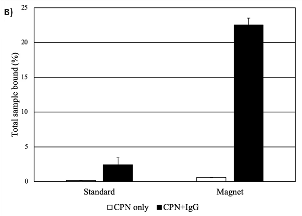
Figure 4. A) Solutions of CPNs under UV illumination and magnetic fields, outlining their dual modality for magnetism and fluorescence. (Edited and reprinted from Howes et al.)2 B) CPNs increase propensity for binding in a plate-based IgG binding assay. CPNs either unlinked or linked to IgG were incubated on plates coated with protein A/G to measure binding events. CPNs linked to IgG display a far stronger assay signal when a magnet is applied, as CPNs are pulled down towards the bottom of the plate, locally increasing their concentration at the site of the target. Image Credit: Merck
Optical Properties
CPNs, with their remarkable brightness and strong photostability, enhance various imaging and diagnostic techniques. The color of light emitted by CPNs depends on the specific polymer core, with products offered in a range of fluorescence emission wavelengths.
Seven visible emission wavelengths are currently available, each with discrete peaks at 420 nm, 435 nm, 475 nm, 510 nm, 550 nm, 610 nm, or 680 nm, along with peaks at 900 nm and 1130 nm (Figure 5).
Studies indicate that conjugated polymer nanoparticles are 175 times brighter than quantum dots and 1400 times brighter than AF488-dex in cellular applications.3,14
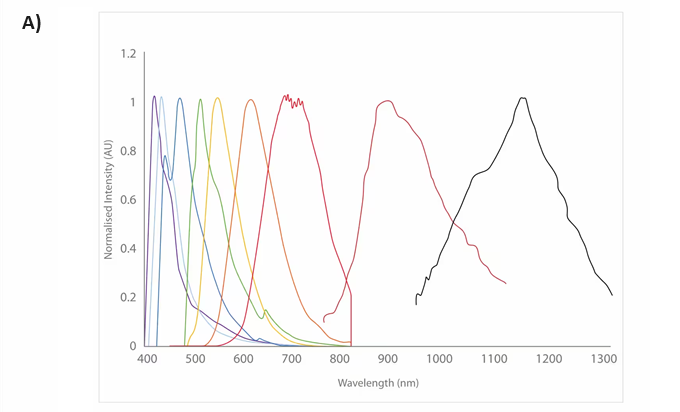
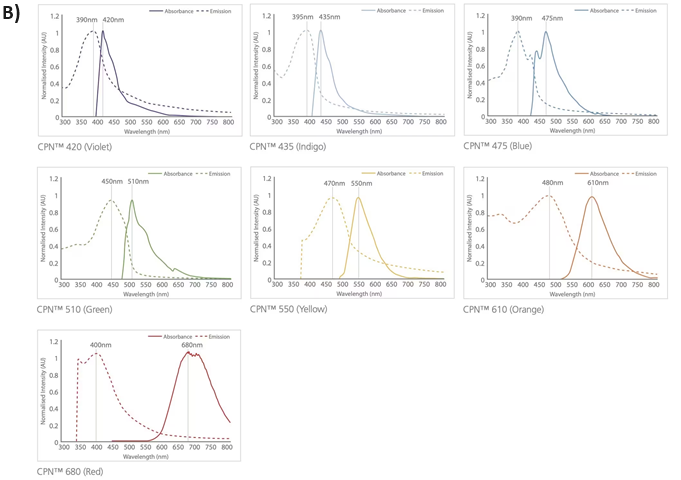
Figure 5. Excitation and emission spectra for the CPN range, covering the visible and NIR spectra. Image Credit: Merck
In both conjugated and non-conjugated states, CPNs maintain their fluorescence for up to 24 months, even under ambient temperature and lighting conditions (Figure 6).
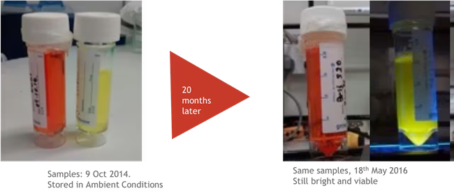
Figure 6. CPNs remain photostable for up to 24 months, retaining their immense fluorescence over a long period of storage. Image Credit: Merck
Enhanced Biofunctionalization
CPNs can label targeted cells through endocytosis or by binding to specific targeting elements like streptavidin, antibodies, or receptor-ligand proteins, serving various binding and targeting applications.
Attaching a CPN to a targeting element involves EDC chemistry (N-ethyl-N'-dimethylaminopropyl-carbodiimide) linking amine groups (-NH2) on the protein to exposed carboxyl groups on the CPN surface (-COOH).
Other surface formulations like thiol and azide cater to click chemistry.
CPNs' brightness ensures highly sensitive detection, enabling the study of individual proteins in samples and cells, detectable even at single molecule levels in flow cytometry and immunocyto/histochemistry.16
Application Versatility
The intense fluorescence, stability, and versatility of CPNs render them remarkably functional and adaptable across a myriad of life sciences applications, enhancing various techniques (Figure 7).
- Fluorescence Microscopy (high content screening, live cell tracking, and 3-D cell imaging)9,16
- Immunohistochemistry and Immunocytochemistry16
- Flow Cytometry5,9,17
- Rapid Diagnostic Tests (RDTs) 12,13
- Tissue Imaging6–8
- 2-Photon Imaging
- NIR Imaging10,11,15
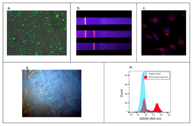
Figure 7. Versatility in a wide range of applications. A) Fluorescence microscopy. HEK293T cell membranes labeled with Cat. No. 905038.9 B) Rapid diagnostic tests with CPN-based rapid diagnostic test strip. Multiple particles can be used simultaneously on one strip for multiple diagnoses.12,13 C) Z-Stack microscopy. HeLa cells stained with Cat. No. 904996 (red) and nuclei stain (DAPI, blue).9 D) Tissue staining using microscopy with UV surface excitation optical section imaging system. Structures can be labeled in paraffin-embedded tissue using Cat. No. 905038 (yellow). E) Flow cytometry. Cat. No. 905038 showing labeling of 50% subpopulation of CD-4 positive cells in flow cytometry.5,9,17 Image Credit: Merck
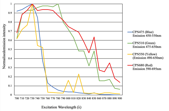
Figure 8. 2-Photon microscopy. Excitation 700–900 nm. Emission CPN 475, Cat. No. 905054 (blue), CPN 510, Cat. No. 905038 (green), CPN 550, Cat. No. 905046 (yellow), CPN 680, Cat. No. 904996 (red). Image Credit: Merck
Summary
CPNs present exciting prospects for fluorescently labeling both molecules and cells. Their extraordinary brightness not only enables the detection and imaging of low-level proteins and rare cell types but also permits the sparing use of lower excitation levels in delicate cells and tissues.3
If necessary, the durability of CPNs allows them to withstand high-intensity illumination and extreme experimental conditions. Their 24-month shelf-life facilitates convenient storage at room temperature. Additionally, they can endure temperatures up to 120 °C and a pH range from 2 to 10.
The unique combination of brightness, stability, magnetic capability, and compatibility with a wide array of molecules positions CPNs to provide substantial enhancements and advantages across a wide spectrum of life sciences applications.
References
- Tuncel D, Demir HV. 2010. Conjugated polymer nanoparticles. Nanoscale. 2(4):484. https://doi.org/10.1039/b9nr00374f
- Howes P, Green M, Bowers A, Parker D, Varma G, Kallumadil M, Hughes M, Warley A, Brain A, Botnar R. 2010. Magnetic Conjugated Polymer Nanoparticles as Bimodal Imaging Agents. J. Am. Chem. Soc.. 132(28):9833-9842. https://doi.org/10.1021/ja1031634
- Fernando LP, Kandel PK, Yu J, McNeill J, Ackroyd PC, Christensen KA. 2010. Mechanism of Cellular Uptake of Highly Fluorescent Conjugated Polymer Nanoparticles. Biomacromolecules. 11(10):2675-2682. https://doi.org/10.1021/bm1007103
- Piwoski H, Michinobu T, Habuchi S. 2017. Controlling photophysical properties of ultrasmall conjugated polymer nanoparticles through polymer chain packing. Nat Commun. 8(1): https://doi.org/10.1038/ncomms15256
- Li K, Liu B. Polymer-encapsulated organic nanoparticles for fluorescence and photoacoustic imaging. Chem. Soc. Rev.. 43(18):6570-6597. https://doi.org/10.1039/c4cs00014e
- Jiang Y, Novoa M, Nongnual T, Powell R, Bruce T, McNeill J. 2017. Improved Superresolution Imaging Using Telegraph Noise in Organic Semiconductor Nanoparticles. Nano Lett.. 17(6):3896-3901. https://doi.org/10.1021/acs.nanolett.7b01440
- Bruns OT, Bischof TS, Harris DK, Franke D, Shi Y, Riedemann L, Bartelt A, Jaworski FB, Carr JA, Rowlands CJ, et al. 2017. Next-generation in vivo optical imaging with short-wave infrared quantum dots. Nat Biomed Eng. 1(4): https://doi.org/10.1038/s41551-017-0056
- Sun H, Zhang Y, Chen Y, Liu Y. 2016. Polyanionic Cyclodextrin Induced Supramolecular Nanoparticle. Sci Rep. 6(1): https://doi.org/10.1038/s41598-016-0026-z
- Chen C, Huang Y, Liou S, Wu P, Kuo S, Chan Y. 2014. Near-Infrared Fluorescent Semiconducting Polymer Dots with High Brightness and Pronounced Effect of Positioning Alkyl Chains on the Comonomers. ACS Appl. Mater. Interfaces. 6(23):21585-21595. https://doi.org/10.1021/am506577r
- Liu H, Wu P, Kuo S, Chen C, Chang E, Wu C, Chan Y. 2015. Quinoxaline-Based Polymer Dots with Ultrabright Red to Near-Infrared Fluorescence for In Vivo Biological Imaging. J. Am. Chem. Soc.. 137(32):10420-10429. https://doi.org/10.1021/jacs.5b06710
- Liu J, Feng G, Ding D, Liu B. 2013. Bright far-red/near-infrared fluorescent conjugated polymer nanoparticles for targeted imaging of HER2-positive cancer cells. Polym. Chem.. 4(16):4326. https://doi.org/10.1039/c3py00605k
- Feng L, Liu L, Lv F, Bazan GC, Wang S. 2014. Preparation and Biofunctionalization of Multicolor Conjugated Polymer Nanoparticles for Imaging and Detection of Tumor Cells. Adv. Mater.. 26(23):3926-3930. https://doi.org/10.1002/adma.201305206
- Li M, Nie C, Feng L, Yuan H, Liu L, Lv F, Wang S. 2014. Conjugated Polymer Nanoparticles for Cell Membrane Imaging. Chem. Asian J.. 9(11):3121-3124. https://doi.org/10.1002/asia.201402711
- Wu C, Szymanski C, Cain Z, McNeill J. 2007. Conjugated Polymer Dots for Multiphoton Fluorescence Imaging. J. Am. Chem. Soc.. 129(43):12904-12905. https://doi.org/10.1021/ja074590d
- Hashim Z, Green M, Chung PH, Suhling K, Protti A, Phinikaridou A, Botnar R, Khanbeigi RA, Thanou M, Dailey LA, et al. Gd-containing conjugated polymer nanoparticles: bimodal nanoparticles for fluorescence and MRI imaging. Nanoscale. 6(14):8376-8386. https://doi.org/10.1039/c4nr01491j
- Debbage P, Jaschke W. 2008. Molecular imaging with nanoparticles: giant roles for dwarf actors. Histochem Cell Biol. 130(5):845-875. https://doi.org/10.1007/s00418-008-0511-y
- Park JH, Oh N. Endocytosis and exocytosis of nanoparticles in mammalian cells. IJN.51. https://doi.org/10.2147/ijn.s26592

This information has been sourced, reviewed and adapted from materials provided by Merck.
For more information on this source, please visit Merck.