Sponsored by NanoImagesReviewed by Olivia FrostJul 22 2025
Raman spectroscopy in a scanning electron microscope (SEM) is a powerful Correlative Light and Electron Microscopy (CLEM) approach that combines electron beam imaging with chemical analysis.

Image Credit: Anucha Cheechang/Shutterstock.com
Raman analysis works by shining a laser on a sample and detecting the inelastically scattered photons. The resulting Raman spectrum provides information about the molecular structure and composition of the material.
Integrating Raman spectroscopy into the electron microscope enables simultaneous high-resolution imaging and chemical analysis of the same area on the sample.
Benefits of Raman in the SEM
Enhanced Analysis Capabilities
Raman spectroscopy in the SEM offers several key advantages: it provides chemical information without the need for sample preparation, works on any non-metal material with molecular vibrations, and does not damage the sample.
Practical Applications
Materials scientists rely on Raman analysis to study geological samples, carbon materials like graphene, semiconductor devices, and pharmaceutical compounds.
Tabletop SEM Integration
Integrating Raman capabilities into a tabletop SEM brings advanced analytical power to smaller laboratories. Previously, achieving this level of analysis required multiple expensive instruments, but now compact systems enable simultaneous imaging and Raman spectroscopy in a single desktop unit.
Combining Raman and SEM analysis in a single instrument saves both money and space. Instead of purchasing separate devices, laboratories can invest in one versatile system.
Applications Across Fields
Materials Science
Materials scientists frequently utilize Raman-enabled SEMs to investigate crystal structure, characterize defects, identify phases, and analyze stress/strain.
Geological Studies
Raman analysis in the SEM aids geologists by enabling the identification of mineral phases, the analysis of meteorite samples, and the examination of gemstones.
Pharmaceutical Research
In the pharmaceutical industry, Raman-SEM systems assist researchers in analyzing and comparing drug formulations, ensuring quality control (including coating uniformity), and identifying contamination.
Technical Considerations
Resolution and Sensitivity
The integration of Raman spectroscopy with the SEM combines the excellent spatial resolution of the SEM with sub-10 µm Raman precision. This allows researchers to directly correlate surface morphology with chemical composition.
Emerging Applications
Looking ahead, scientists expect Raman-SEM systems to play a growing role in nanotechnology, battery research, biological materials, and environmental studies.
Image Credit: NanoImages
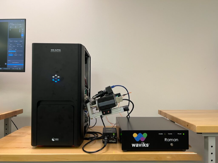
Image Credit: NanoImages
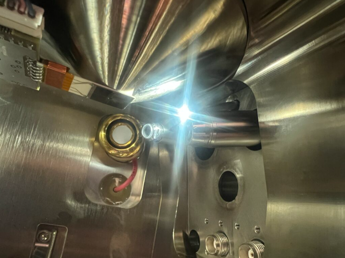
Image Credit: NanoImages
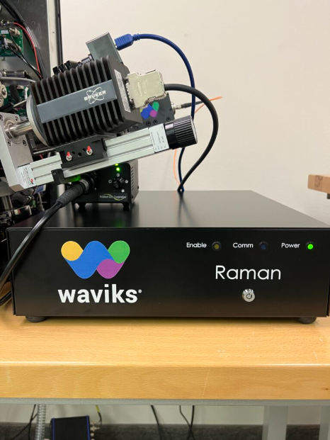
Image Credit: NanoImages
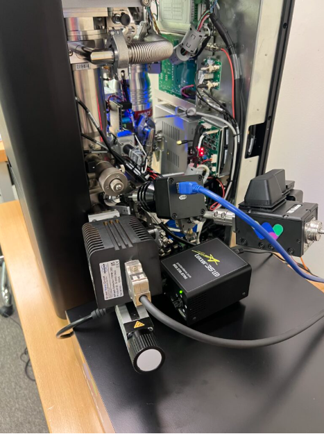
Image Credit: NanoImages
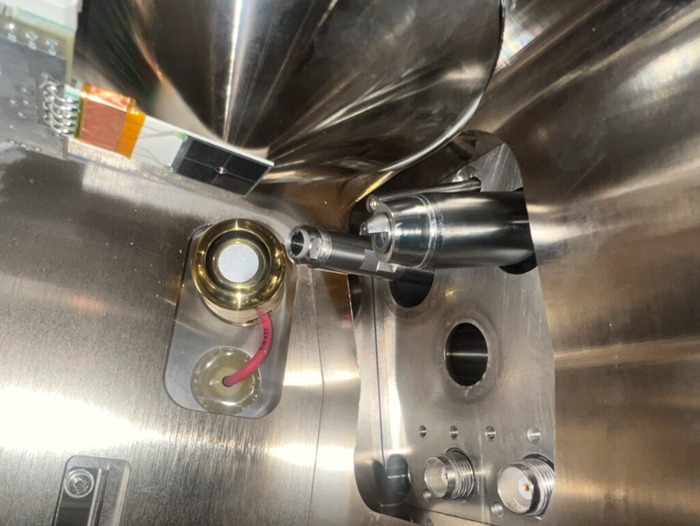
Image Credit: NanoImages
Image Credit: NanoImages
Conclusion
Raman spectroscopy in a tabletop SEM combines high-resolution imaging with powerful chemical analysis in a format that is more accessible to smaller laboratories and mobile workshops.

This information has been sourced, reviewed and adapted from materials provided by NanoImages.
For more information on this source, please visit NanoImages.