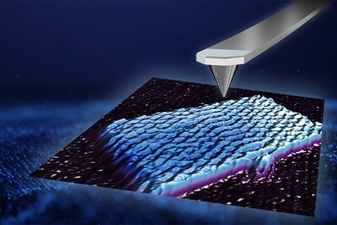
In this interview, AZoNano speaks with Dr. George Heath from the University of Leeds, UK, about the fundamental principles of Atomic Force Microscopy (AFM), integration with advanced optical microscopy, and relevant applications. He also discusses his research utilizing the NanoRacer high-speed atomic force microscope to examine protein dynamics. He also looks at techniques that enhance AFM's temporal and spatial resolution down to 10 microseconds and sub-nanometer levels.
What makes high-speed AFM valuable for studying dynamic biological systems?
High-speed AFM is particularly valuable for studying dynamic biological systems because it allows us to capture rare and stochastic structural changes in proteins and biomolecules with unprecedented detail.
Unlike other techniques that may lack the necessary resolution to observe these changes, high-speed AFM provides high spatial and temporal resolution, enabling us to see individual molecules and their interactions in real-time.
This is crucial for understanding complex biological processes, such as protein-protein interactions, membrane dynamics, and conformational changes, which are often driven by rare events and occur on short timescales. High-speed AFM allows us to track these events as they happen, gaining insights into the mechanisms underlying biological function.
How do advancements in high-speed AFM, particularly with the NanoRacer, address the limitations of standard AFM in terms of spatial and temporal resolution?
Advancements in high-speed AFM, particularly with the NanoRacer, address the limitations of standard AFM by significantly improving spatial and temporal resolution. Traditional AFM techniques often struggle with slow scanning speeds and limited resolution, making it challenging to observe dynamic processes in real-time. However, with the NanoRacer and other high-speed AFM systems, we can overcome these limitations.
Using smaller cantilevers, higher frequency tapping, and better feedback controls, we can achieve scanning speeds of up to 55 frames per second or even higher.
This allows us to capture rapid structural changes with unprecedented detail, providing insights into biological processes that were previously inaccessible. The ability to perform line scanning and localization AFM further enhances the spatial resolution of AFM images, allowing us to observe molecular interactions at the single-molecule level.
High-Speed Atomic Force Microscopy - DNA imaging at 50 frames per second
Bruker JPK NanoRacer® high speed AFM video of part of individual DNA molecule imaged in fluid on mica+PLO substrate in closed-loop, the video contains 1000 images acquired without user assistance at an imaging speed of 50 frames/sec demonstrating the stability and low force imaging of the NanoRacer® AFM at these high speeds, x-range 100 nm (100 pixels), y-range 80 nm (80 pixels), z-range 2.0nm. Image Credit: Bruker BioAFM
Can you provide examples of dynamic biological processes successfully studied using high-speed AFM, particularly with the NanoRacer?
High-speed AFM has been instrumental in studying various dynamic biological processes. One notable study involved observing the dynamics of Myosin V motor protein walking along actin filaments. This study revealed the stepping mechanism of Myosin V, providing insights into how motor proteins move and interact with their cellular environment.
We have also studied the behavior of antimicrobial peptides attacking bacterial membranes and the binding and unbinding kinetics of proteins to lipid membranes. These examples demonstrate the versatility of high-speed AFM in studying a wide range of dynamic biological processes with high spatial and temporal resolution.

Image Credit: Bruker BioAFM
How does high-speed AFM enable the observation of protein-protein interactions and conformational changes at the single-molecule level?
High-speed AFM enables the observation of protein-protein interactions and conformational changes at the single-molecule level by capturing these events in real-time with high precision.
With the NanoRacer, we can track the interactions between individual protein molecules as they occur, allowing us to observe binding and unbinding events, as well as conformational changes, with nanometer-scale resolution.
For example, we have been able to study the conformational changes of Annexin V protein as it binds to lipid membranes in the presence of calcium ions. By imaging the protein at high speeds, we can observe how it interacts with the membrane and undergoes structural changes in response to the binding process.
You developed a new image analysis tool, NanoLocz. Could you explain how it handles the extensive data generated by high-speed AFM observations?
NanoLocz, our freely available new image analysis tool, addresses the challenge of handling extensive data generated by high-speed AFM observations through several key features. First, it allows for easy data import by supporting all AFM file formats, ensuring compatibility with various high-speed AFM systems.
Once the data is imported, NanoLocz employs parallel processing techniques to expedite pre-processing and analysis, enabling it to handle multiple images simultaneously. This significantly reduces the time required for data processing.
One of NanoLocz's functions is automated leveling, which ensures that AFM images are properly flattened and aligned, enhancing the accuracy of subsequent analyses. The software also includes advanced particle detection and super-resolution algorithms, enabling it to identify and improve the resolution of individual molecules, protein complexes, or other structures, even within crowded datasets.
Could you describe a specific study or experiment where high-speed AFM with the NanoRacer provided novel insights into the dynamics of a biological system that were previously unattainable with other techniques?
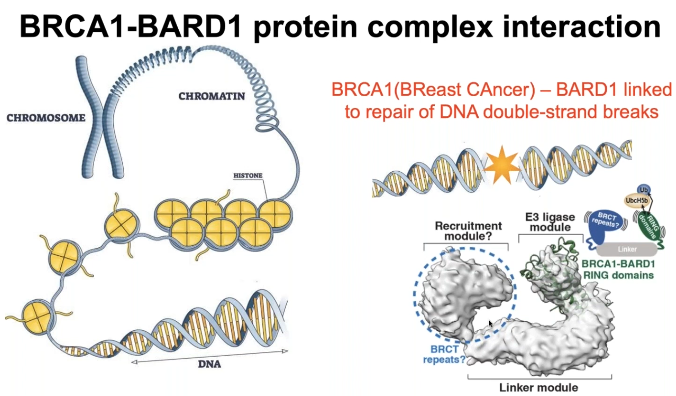
One notable study where high-speed AFM with the NanoRacer provided novel insights into a biological system's dynamics is the investigation of the BRC-BARD1 protein complex involved in DNA repair. This study aimed to understand how this complex interacts with DNA at the molecular level, which is crucial for our understanding of DNA repair mechanisms and their implications in cancer.
Traditionally, the structure of the BRCA1-BARD1 complex was challenging to elucidate using techniques like cryo-electron microscopy (cryo-EM) or X-Ray crystallography due to its inherent flexibility. However, high-speed AFM with the NanoRacer allowed researchers to directly visualize the dynamic interactions between BRCA1-BARD1 and DNA in real-time.
Using high-speed AFM, researchers observed that the BRCA1-BARD1 complex could bind to individual nucleosomes and bridge across multiple nucleosomes, a previously unattainable phenomenon with other techniques.
This finding provided crucial insights into the mechanism by which BRCA1-BARD1 facilitates DNA repair. It suggests that the complex can stabilize DNA structures across multiple nucleosomes, thereby promoting efficient repair processes.

Image Credit: George Heath
How do line scanning and localization AFM improve the time and spatial resolution of AFM images, and how do these techniques enhance our understanding of dynamic biological systems?
Line scanning involves repeatedly scanning a single line across an area of interest. It allows researchers to achieve higher time resolution by focusing on specific regions where dynamic events are occurring. By limiting the scanning area, line scanning reduces the time required to capture images, enabling the observation of rapid biological processes with millisecond or even microsecond resolution.
This technique enhances our ability to track dynamic events such as protein conformational changes or molecular interactions in real time, providing valuable insights into the kinetics and mechanisms of biological processes.
Localization AFM improves spatial resolution by localizing peaks in AFM data and reconstructing images from these localized points. This technique enables researchers to achieve resolutions beyond the limits of traditional AFM imaging, approaching the scale of individual atoms or molecular features. By precisely pinpointing the positions of fluctuations within molecules or protein complexes, localization AFM allows for the visualization of fine structural details and the characterization of molecular interactions with unparalleled accuracy.
By combining line scanning and localization AFM, researchers can observe dynamic biological processes at both high-time and spatial resolutions. This enables the visualization of molecular movements, conformational changes, and interactions with unprecedented detail, shedding light on the underlying mechanisms of biological function.
What are the potential future directions for advancements in high-speed AFM technology, and how might they impact the study of dynamic biological systems?
The potential future directions for advancements in high-speed AFM technology are vast and promising, with several key areas likely to impact the study of dynamic biological systems.
One direction involves further improving the speed and sensitivity of high-speed AFM systems. This could involve innovations in cantilever design, feedback mechanisms, and detection methods to enable even faster imaging speeds and higher sensitivity. By increasing the temporal resolution of high-speed AFM, researchers can capture even more rapid biological processes with greater detail, allowing for the study of dynamic events that were previously inaccessible.
Another area of advancement is enhancing the versatility and capabilities of high-speed AFM for studying complex biological systems. This could include developing new imaging modes and techniques tailored to specific biological questions, such as imaging at the interface of living cells.
Integration with other imaging modalities is also a promising direction for future advancements in high-speed AFM technology. Combining AFM with techniques such as fluorescence, super-resolution, or electron microscopy could provide complementary information about dynamic biological processes, allowing researchers to correlate structural and functional changes at multiple scales.
About Dr. George Heath
George Heath is a University Academic Fellow specializing in experimental biophysics with a focus on high-speed atomic force microscopy (AFM). His academic and professional journey, marked by an impressive blend of research and development in physics and life sciences, underscores his commitment to advancing our understanding of biological systems at the molecular level.
George pursued his higher education at the University of Leeds, where he obtained both his Master of Physics (MPhys) and Doctor of Philosophy (Ph.D.) in Physics. Following his Ph.D., George continued at the University of Leeds as a Postdoctoral Fellow from 2014 to 2017. In July 2017, George expanded his research horizon by joining Weill Cornell Medicine in New York as a Postdoctoral Associate for two years. In 2019, he returned to the University of Leeds to start his own research group, where he is currently an EPSRC Open Fellow appointed across the School of Physics & Astronomy and the School of Biomedical Sciences.
An active contributor to the scientific community, George's publications reflect his innovative approach to biophysics. His notable works include the development of Localization AFM, AFM Height Spectroscopy and several studies of protein and membrane dynamics. These contributions not only underscore his expertise but also his commitment to pushing the boundaries of what's possible in biophysical research.

This information has been sourced, reviewed and adapted from materials provided by Bruker BioAFM.
For more information on this source, please visit Bruker BioAFM
Disclaimer: The views expressed here are those of the interviewee and do not necessarily represent the views of AZoM.com Limited (T/A) AZoNetwork, the owner and operator of this website. This disclaimer forms part of the Terms and Conditions of use of this website.