With the rapid development of engineered nanoparticles and nanoscale structure-based products and materials, awareness of the poor understanding of potential toxic effects and environmental impact of these materials has also increased. In this regard, current methods have been investigated, and new methods are required to analyze the materials regularly during production and development.
Nanoparticle Tracking Analysis, or NTA, is a versatile and rapid multi-parameter technique designed for the characterization of nanoparticles. The ability to provide number frequency particle size distributions of polydisperse nanoparticulate systems has made this method suitable for environmental and toxicity studies in various sectors. This article describes some of the recent works carried out by researchers to investigate nanoparticle toxicity and environmental impact using NTA.
Nanoparticle Tracking Analysis
NTA is operated based on Brownian motion and light scattering properties to provide particle size distribution of samples in liquid suspension. A laser beam is irradiated on a sample chamber containing the sample. The light beam is scattered by the particles in the liquid suspension that come across the light path, and the scattered light can be viewed using a 20x magnification microscope mounted with a camera. Operating at about 30 fps, the camera tracks the particle movement under Brownian motion in nearly 100 x 80 x 10 µm field of view, as shown in Figure 1. The particle movement is captured in a video format.
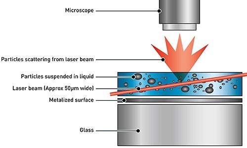
Figure 1. Schematic of the optical configuration used in NTA.
The particle movement is captured on frame-by-frame format. The center of each particle is identified and captured using the proprietary NTA software. The software also evaluates the average distance covered by the particle in both the x and y planes. Based on this value, the particle diffusion coefficient (Dt) can be determined. Further, using the Stokes-Einstein equation as given below, the sphere-equivalent hydrodynamic diameter, d, of the particles can be calculated with known sample temperature T, solvent viscosity η and Dt.
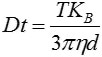
Figure 2 shows an example of the size distribution profile created by NTA. The NTA can also be used to measure the particle movement in a fixed field of view that is illuminated by a 10 µm deep beam. The scattering volume of the sample can also be estimated from the figures.
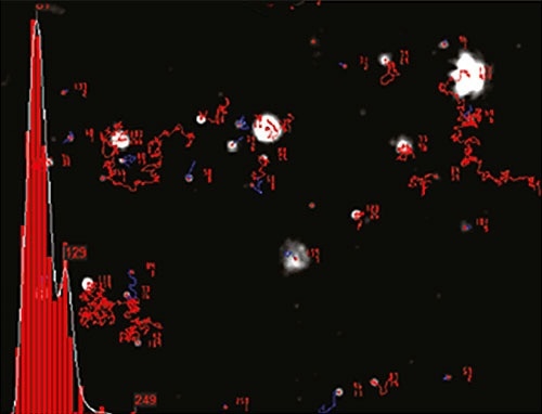
Figure 2. An example of the size distribution profile generated by NTA. The modal size for this sample is found to be approximately 70 nm, with larger sized particles also present.
Cytotoxic Studies
NTA was used in the investigation of the genotoxicity of cobalt nanoparticles in human peripheral leukocytes (Colognato et al. 2008) and mouse fibroblasts (Ponti et al. 2009) at cellular level. Further, with this method, Wick et al. (2009) reported the penetration of nanoparticles via the human placenta and also the transport of SiO2 nanoparticles via human skin (Staronova et al., 2012). Filton et al. (2012) showed that cobalt nanoparticles can penetrate the human skin via intact and damaged skin, suggesting the use of cobalt nanoparticles in an in-vitro diffusion cell system.
A number of studies focus on the potential impact of different metals on a number of cellular and aqueous systems using NTA, including the effect of copper and chrome oxide nanoparticles (Studer et al. 2010, Khatoonet al. 2011), silver nanoparticles (MacCuspie et al. 2011, Bouwmeester et al. 2011) and gold nanoparticles (Gosens et al. 2010).
Knowledge on the nanoparticle’s size dispersion distribution is critical for their penetration into cellular systems to their cytotoxicity. In this aspect, NTA has proved successful over other nanoparticle characterization techniques including Dynamic Light Scattering (DLS) (Kendall et al. 2009, Patel et al. 2010, Munaro 2010, Karlsson 2010).
Using NTA, Christen and Fent (2012) reported the initiation of endoplasmatic reticulum stress response and variation of cytochrome P4501A activity in human liver cells (Huh7) and Pimephales promelas (fathead minnow) fibroblast cells (FMH) followed by the injection of engineered silica nanoparticles and silver-doped silica nanoparticles. The NTA was used to study the particle’s stability in nanopure water. Choudhary et al. (2013) investigated the impact of cigarette smoke on PA pressures and ventricular function by NTA.
The cell behavior is greatly influenced by the physicochemical properties of nanoparticles. However, the effect can be minimized by the protein-related interactions in biological solutions. Recently, Bartczak et al. (2013) used NTA to illustrate the size distribution changes and nanoparticle dissolution in serum containing cell culture media, which are caused by the different surface functionalities of zinc oxide (ZnO) nanoparticles.
Aquatic and Marine Toxicity
NTA technique could also be used to study the aquatic environmental impact and potential cellular toxicity of nanoparticles in the future (Hassellöv and Kaegi, 2009). This study explained the challenge in modeling and quantifying the complex interactions between engineered nanoparticles and aquatic environmental matrices, where NTA can be employed.
Further, a number of research programs explored the ecotoxicological effects of various nanoparticles, for instance on the freshwater environment (Juhel 2009). The effects of silver and gold nanoparticles on fish (Scown et al., 2010), rainbow trout hepatocytes and gill cells (Farkas et al., 2010, Farkas et al., 2011) as well as the effects of CeO2 that serves as a catalyst in the automotive industry (Quik et al., 2010; Van Hoecke et al,. 2009) were also studied.
Trumsina carried out a comparative analysis on different methods used for monitoring the separation of nanoparticle from textiles during washing, and provided the significant benefits of both NTA and DLS over a new gas discharge visualization method.
NTA has also played a major role in probing the effect of soluble (FeCl3) iron, and engineered Fe2O3 nanoparticles on the developmental toxicity due to CO2-induced seawater acidification (Kadar et al. 2010). In a recent study, Tatarkiewicz et al. (2012) discussed the role of NTA in measuring concentration and sizing of colloidal particles in the Arctic Ocean. Similarly, Stuart et al. (2012) described the proof-of-concept measurements on the nanoparticles effect over an electrode potentiostatted at a value, with respect to the diffusion controlled oxidation of silver nanoparticles in authentic seawater media.
Microbiota and Plants
NTA has proved useful in studying the impact of nanoparticles on microorganisms and their ecology. It helps in proving information related to the properties and behavior of nanoparticulates. In 2011, Kroll and her Co-workers used a number of methods including NTA to investigate the distribution and bioavailability of engineered nanoparticles such as titanium dioxide, cerium dioxide and silver nanoparticles in freshwater periphyton. They evaluated the material properties including dissolution, charge and size.
Turner et al. (2011) carried out a similar investigation on the interactions of Ag nanoparticles with Ulva lactuca, marine microalgae using NTA in order to characterize their Ag nanoparticle suspensions. Chaudhari et al. (2012) studied the influence of biosynthesized silver nanoparticles on Staphylococcus aureus biofilm quenching and prevention of biofilm formation using electron dispersive X-ray spectra, NTA and TEM.
NTA has also been reported in bacterial research studies. Park et al. (2012) assessed the antibacterial and water purification properties of self-assembled honeycomb structure of aerosol deposited titania film. Similarly, Khatoon et al. (2011) used NTA to determine the internalization of chromium oxide nanoparticles in Escherichia coli by flow cytometry. Carter et al. (2012) reported significant reduction in Escherichia coli O157:H7 contamination of lettuce and beef using a bacteriophage cocktail which, however failed to prevent recontamination.
Kadar et al. (2012) studied the impact of coatings on the zero-valent nano-iron (nZVI) on early life stages of three important marine invertebrate species. They used NTA to monitor the dissolution of nZVI in sea water to show how the coating helps in stabilizing the nanometal suspension. This was followed by the study on effects of NTA-analyzed industrially relevant engineered iron nanoparticles on growth and metabolic status of marine microalgae cultures.
NTA has been central to studies related to the transport of nanoparticulates in fungi. Cunha-Azevedo developed and investigated an anti-fungal formulation based on PLGA nanoparticles that release active agent itraconazole. The findings revealed the size as an important parameter to be considered, which when analyzed by NTA was found to be average of 174nm (Cunha-Azevedo 2011).
Hartmann et al. (2012) analyzed the challenges involved in testing metal and metal oxide nanoparticles in algal bioassays with the help of titanium dioxide and gold nanoparticles. They used NTA to determine the hydrodynamic diameter and size distributions of suspended particles.
Piccapietra et al. (2011) measured variations in the physicochemical properties of silver nanoparticles (AgNP) while studying the bioavailability, mobility and fate of AgNP in aquatic systems, using NTA, DLS and ultraviolet-visible spectroscopy. Recently, Matzke et al. (2013a) discussed the effects of selected silver nanoparticles on freshwater microbial species using NTA to determine particle size distribution.
While studying the effect of Ag and Al2O3 nanoparticles on the soil bacteria, Bacillus cereus and Pseudomonas stutzeri using NTA, Fajardo et al. (2013) proved the presence of bactericidal effects on exposure to 5 mg/L Ag nanoparticles for 48 hour. The findings also suggested that less or no toxicity was caused by Al2O3 nanoparticles while assaying. Zhdanov and Hook (2013) showed the nucleation in mesoscopic systems in transient conditions corresponding to the peptide-induced pore formation in vesicles.
Vajpayee et al (2011) used NTA to study nanoparticulates transport in seed germination. The method has also been used to investigate nanoparticulates transport in surface runoff across dense vegetation (Yu, 2011).

This information has been sourced, reviewed and adapted from materials provided by Malvern Panalytical.
For more information please visit Malvern Panalytical.