Discover the effortless integration process that demands minimal effort and delivers exceptional user experience.
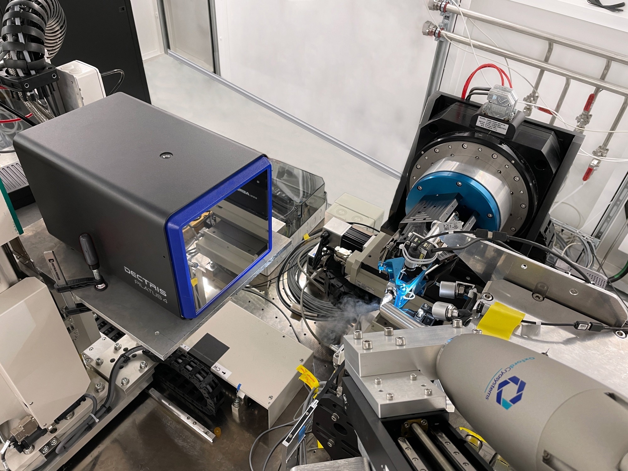
A DECTRIS PILATUS4 CdTe 1M detector prototype at the Swiss Light Source (SLS). Image Credit: DECTRIS AG
At the SLS PXIII beamline, a DECTRIS PILATUS4 CdTe 1M detector prototype was tested for protein crystallography. The goal was to assess the suitability of the detector for the beamline following the upgrade of the SLS synchrotron to SLS 2.0, which had only just begun.
The final two weeks of SLS’ operation could be used efficiently for testing, and even for collecting datasets - all thanks to the fast and smooth integration of the detector.
Members of the SLS’ Macromolecular Crystallography (MX) group were recently asked about their experience with DECTRIS.
How did you integrate PILATUS4 into your beamline control system?
Ezequiel: The integration of the PILATUS4 detector into the beamline control system was a simple process because the platform comes equipped with the SIMPLON interface, the same one we use to control the EIGER detectors (the data and control interface SIMPLON API). This enabled us to make better use of our existing Python modules and seamlessly control the PILATUS4 detector.
It was remarkably easy to integrate, as we could configure the detector and initiate a data acquisition series in just minutes of it being connected to the network. Having previously worked on integrating a variety of detectors, this was one of the smoothest we’ve ever conducted.
Wayne: It was initially assumed that a significant amount of effort would be required. We were pleasantly surprised that the process went incredibly smoothly and was efficiently executed.
Ezequiel: The immediate access to streamed images for our data analysis software when conducting grid scans is also worth noting. This was of great use, as it eliminated the need to make adjustments to the existing software infrastructure. The entire process was highly efficient and user-friendly, as the data generated by the PILATUS4 detector was seamlessly compatible with our analysis tools. This is a significant advantage when it comes to data accessibility and compatibility.
Could you describe the amount of effort required to get the detector up and running, from when it was delivered to when the first scientifically usable dataset was acquired?
Ezequiel: I would say it definitively required minimal effort - essentially zero. The process was very smooth. Setup began in the morning, and by just after lunchtime, the system was basically up and running.
Identifying the MAC address on our network and dealing with the required 802.1x authentication was the most challenging part, and that took approximately 3 minutes.
Did you come across any particular challenges?
Kate: The process was quite streamlined, and we didn’t face any notable issues. It worked seamlessly ‘out of the box’ with the X-Ray Detector Software in terms of automatic data processing. The process of configuring the detector’s dimensions in the input file was straightforward, needing only a simple click. It was very much a user-friendly experience.
What was the maximum speed at which you collected data with the PILATUS4, and what do you expect in terms of speed once the upgraded SLS 2.0 synchrotron is operational?
Meitian: We achieved data collection speeds of up to 500 Hz with the PILATUS4. In practice, 100-200 Hz is an acceptable rate for daily operations, but specific projects may require an additional few hundred Hz.
Wayne: With significantly higher flux after the SLS upgrade, we could possibly push the data collection speed higher, with the aim of a maximum of 2,000 Hz in extreme scenarios.
What is your assessment of using a pixel size of 150 µm for protein crystallography?
Vincent: At this time, spot separation is not a large concern for us, generally because we do not deal with large Unit Cells that require extensive separation and longer. In terms of fragment screening, the spot separation process is usually straightforward and works with well-characterized proteins. In truth, a large portion of our crystal samples that are brought to synchrotrons and beamlines land in this category.
Wayne: An advantage of making use of larger pixels (compared to those of EIGER) is a reduced number of pixels that are needed to cover a surface, resulting in faster data processing pipelines and less storage space. That being said, it’s vital to verify whether similar data quality can be achieved with fewer pixels. This assessment should be based on data quality considerations independent of the pixel size.
Related to the larger pixel size, can you solve the structure effectively and observe high-resolution details such as ligands?
Vincent: Definitely. This can result in accelerated data processing and improved efficiency in data transfer downstream.
After you have utilized the 1M-pixel CdTe detector for your tests, what type of detector would you consider the optimal choice for your beamline?
Kate: Our preference remains with silicon as the ideal choice because it continues to perform effectively, even at lower energies. We still require the ability to extract high-quality data at lower energies, which makes silicon a great option for our needs.
Do you recommend the PILATUS4 detector for Macromolecular Crystallography? If so, for which specific applications?
Vincent: It is highly recommended for conventional crystallography work, such as regular cryo-measurement and fragment screening. In these applications, it performs very well.
With the upgrade to SLS 2.0 and a PILATUS4 detector, what is the maximum resolution you would anticipate achieving for protein crystallography?
Meitian: Our goal is a resolution of around 1.5 Å for protein crystallography, which is probably the upper limit. In other applications, such as chemical crystallography, the target resolution is approximately 0.7 Å. These applications require the use of higher X-Ray energy and a larger detector, e.g., a PILATUS4 2M.
About the speakers
Dr. Meitian Wang
Dr. Meitian Wang is currently serving as a Group Leader and Beamline Scientist at the Swiss Light Source (SLS) located at The Paul Scherrer Institute (PSI) in Switzerland. Having obtained a Ph.D. from the University of Alberta in Edmonton, Canada, Dr. Wang specializes in advancing instrumentation and methodology within macromolecular crystallography (MX) beamlines, contributing to research efforts in both academic and proprietary domains.
Wayne Glettig
Wayne Glettig has been a member of The Paul Scherrer Institute (PSI) team since 2017, initially serving as a beamline engineer and subsequently assuming the role of Group Leader for the MX Instrumentation Group in 2018. With a master's degree in microengineering obtained from EPF in Lausanne in 2006, Wayne’s career has encompassed significant contributions to scientific instrumentation, precision mechanisms, and robotics, spanning roles at EPFL, PSI's MX group, and CSEM in Neuchâtel.
 Dr. Vincent Olieric
Dr. Olieric holds the position of Beamline Scientist at the Swiss Light Source (SLS) within the Paul Scherrer Institute (PSI). He pursued a biochemistry degree at the University of Strasbourg and completed his undergraduate studies at Lehigh University in Pennsylvania, USA. Following a master's in Crystallography, he obtained a Ph.D. in Structural Biology in 2006. With extensive experience in synchrotron beamline instrumentation development, crystallographic data collection strategies, and structural biology collaborations, Dr. Olieric plays a vital role in beamline operation, upgrades, user support, and advancing structural biology projects at the SLS.
Dr. Kate Mary Louise Smith
Dr. Smith completed her undergraduate studies in Biochemistry and Mathematics at Charles Sturt University, Australia, and continued to excel with a First Class Honours degree in Biochemistry and Structural Biology from the sa me institution. In 2019, she completed her PhD on nuclear import receptors and their interactions with viral proteins and breast cancer-related proteins. She then gained experience as a macromolecular crystallography beamline scientist at the Australian Synchrotron. Dr. Smith joined the Swiss Light Source (SLS) in 2020 and has been an integral member of the MX Data Group contributing to data acquisition and processing software for all three MX beamlines.
Dr. Ezequiel Panepucci
Dr. Panepucci completed his undergraduate studies with a Chemistry degree at the University of São Paulo. Following his Ph.D. research, he transitioned into a career as a Scientific Software developer and Systems Administrator at Yale University's Howard Hughes Medical Institute laboratory, later relocating to Stanford University. Dr. Panepucci then ventured to Paris for a post-doctoral fellowship at the Pasteur Institute. Here, he discovered the protein crystallography beamlines at the Swiss Light Source (SLS), where he found his true calling as a software developer for the Macromolecular Crystallography beamlines. Today, Dr. Panepucci is dedicated to the development of data acquisition software and its user interface to ensure a productive and enjoyable experience for guest scientists conducting cutting-edge research at the SLS facilities.

This information has been sourced, reviewed and adapted from materials provided by Dectris Ltd.
For more information on this source, please visit Dectris Ltd.
Disclaimer: The views expressed here are those of the interviewee and do not necessarily represent the views of AZoM.com Limited (T/A) AZoNetwork, the owner and operator of this website. This disclaimer forms part of the Terms and Conditions of use of this website.