AFM-IR refers to a broad category of nanoscale chemical characterization techniques that combine Atomic Force Microscopy (AFM) with infrared (IR) spectroscopy. The goal is to generate high-resolution IR spectra and absorption maps at spatial resolutions beyond the diffraction limit of infrared light. For this reason, the term "nano-IR" is often used interchangeably with AFM-IR.
This approach enables detailed analysis of a wide variety of samples, including complex nanocomposites and nanoparticles. AFM is well-suited for these applications because it requires minimal sample preparation. Additionally, the AFM tip plays a key role in bypassing the traditional resolution limits of infrared light through innovative detection methods.
AFM Background
AFM is a scanning probe technique that offers exceptionally high-resolution images of a sample surface. The most advanced AFMs approach atomic resolution, though this typically requires advanced techniques and specialized environments like ultra-high vacuum.
Typical resolutions are less than 10 nm in ambient conditions, well below the diffraction limit of visible or infrared light.
An AFM probe operates similarly to the needle of a record player: while the record player’s needle produces sound through deflection, the AFM’s sharp tip detects surface topography through its deflections. A topographical image is produced by raster scanning the AFM tip over the sample surface.
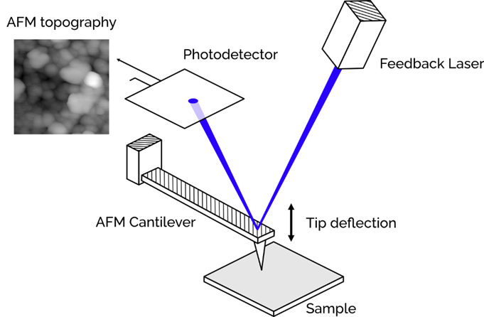
Figure 1. The tip of the AFM "feels" the surface of the sample. The topography is recorded by observing the deflection of the cantilever using a laser feedback system. Image Credit: Molecular Vista
AFM images are created by 'feeling' the sample. The raw data captured is presented as height maps, with black representing low areas and white representing high ones. These can be rendered into three-dimensional surfaces that can be rotated to emphasize key topographical features.
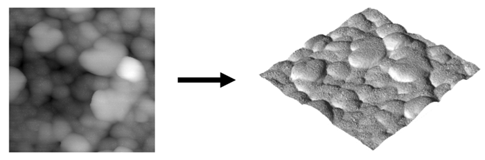
Figure 2. AFM images are essentially height maps that can be used to render the 3D topography. Image Credit: Molecular Vista
AFMs offer several practical advantages. Firstly, virtually any reasonably flat sample can be imaged almost immediately, with no special preparation required.
AFM imaging is also non-destructive when operated in non-contact mode, ensuring the sample remains unaltered during scanning. These benefits make AFM a popular and effective imaging technique, especially when enhanced with infrared capabilities.
Spectroscopy Background
Spectroscopy can be applied in numerous contexts, from studying color shifts in distant stars to analyzing atomic energy transitions.
The most relevant spectroscopy method for AFM-IR is Fourier transform infrared spectroscopy (FTIR), which probes molecular vibrational states to examine the types of bonds in a compound.
FTIR is widely used to study bond structure changes or to identify unknown materials based on their unique spectral signatures. Its broad utility has made FTIR nearly synonymous with chemical characterization.
The advantages of AFM-IR become clearer when considering how it combines the molecular insights of FTIR with the imaging precision and spatial resolution of AFM.
Uses of AFM-IR
AFM-IR techniques occupy a unique middle ground between spectroscopy and microscopy. As a result, this field represents a growing area of specialized expertise. Researchers or technicians already well-versed in either microscopy or spectroscopy may need to adapt their approach when working with AFM-IR methods.
For example, a chemist experienced in IR spectroscopy understands the strengths of techniques like FTIR for complex chemical analysis. IR spectra can reveal changes in bonds or materials within a sample by highlighting characteristic peaks associated with specific bonding combinations.
For these reasons, FTIR plays an important role in forensic, failure, and investigative analysis, offering a way to identify materials based on their unique IR spectra. However, FTIR requires relatively large and thick samples, and the resulting spectra reflect an average of all the nanomaterials present in the sample.
This is where a shift in perspective is needed for chemists familiar with FTIR who are exploring AFM-IR techniques. Nano-IR imaging and spectroscopy make it possible to spatially isolate chemical changes on a sample’s surface with remarkable resolution.
While the potential may be immediately clear to some, consider this analogy to illustrate the difference: think of an RGB LCD display showing a yellow object. Since RGB devices can't emit yellow light directly, they simulate it by blending red and green light using pixels that are too small for the eye to distinguish. With enough magnification, though, you can see the individual pixels.
The shift from FTIR to AFM-IR is similar—Nano-IR data can “see” the individual pixels that FTIR cannot.
While an FTIR spectrometer typically records an aggregate spectrum from a complex sample, AFM-IR can isolate individual spectra and map their precise locations.
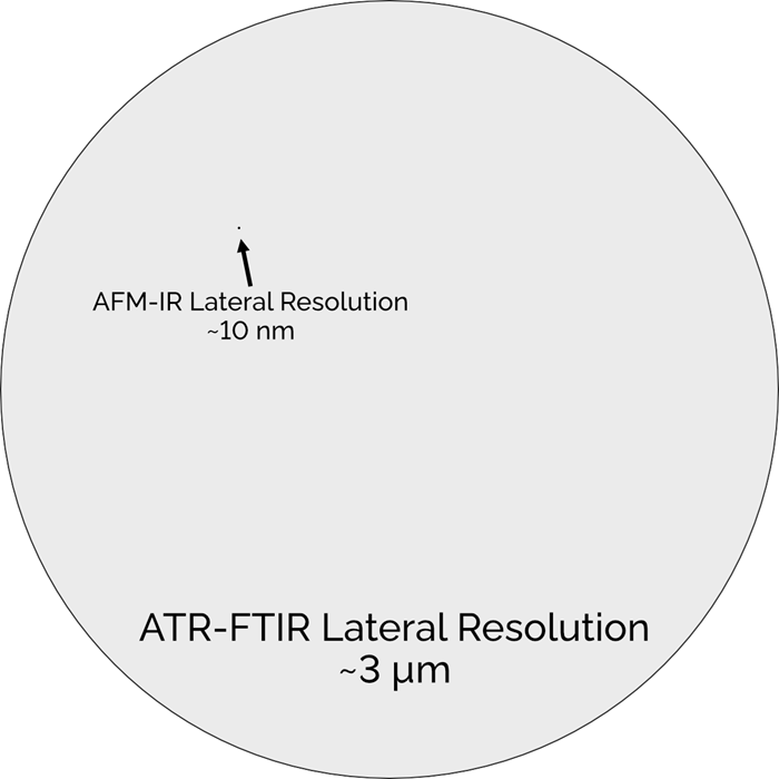
Figure 3. The lateral resolution of AFM-IR techniques is so much higher than FTIR, that it can isolate and probe individual chemical components at the nanoscale. Image Credit: Molecular Vista
This means that when analyzing materials that are truly homogeneous at the nanoscale, spectra from nano-IR techniques often closely resemble FTIR data of the same material.
However, many materials exhibit some degree of inhomogeneity at such small scales, and nano-IR has the resolution to detect those differences.
For studying complex and varied surface chemistry, there’s no better tool than nano-IR imaging. The results may not always align with what’s observed at larger scales, but that’s because factors like molecular orientation or phase come into play.
When the probe measures the response from just a few molecules, it offers an extraordinary view into chemical behavior that can't be captured any other way.
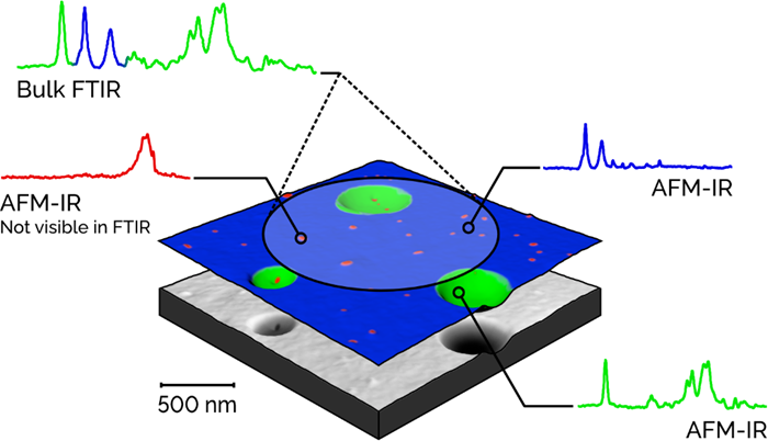
Figure 4. AFM-IR techniques open new possibilities for chemical analysis by providing the resolution necessary to explore single layer chemical films, complex nano-composite materials, nanoparticles, and more. Image Credit: Molecular Vista
For microscopists, the transition to AFM-IR presents a different kind of learning curve compared to spectroscopists. They’re already skilled at examining samples at the smallest possible scales, and those familiar with AFM are used to interpreting scanning probe images and recognizing common artifacts.
However, one ongoing challenge in microscopy is identifying the composition of features within an image, often relying on inference or guesswork.
AFM-IR changes that by enabling microscopists to overlay chemical absorption maps onto their data and collect point-specific spectra to identify unknown features directly. This makes AFM-IR a valuable tool for tasks like defect analysis in semiconductor masks, identifying biomineral inclusions, or pinpointing contaminants.
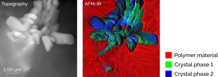
Figure 5. AFM-IR allows microscopists to colorize images with detailed chemical information. Here is a sample with two materials—one of which is a crystal with two different phases. Image Credit: Molecular Vista
AFM-IR’s cross-disciplinary nature requires some adjustment for users with in-depth knowledge of other disciplines, but the power afforded by AFM-IR’s hybrid nature makes it capable of analyses that would be otherwise impossible.
Specific Techniques
Several techniques fall under the umbrella of AFM-IR, with the most widely used being nano-FTIR, tapping mode photothermal IR (TM PTIR), and photo-induced force microscopy (PiFM).
As the developer of PiFM, the first and only non-contact (and therefore non-destructive) AFM-IR technique, Molecular Vista positions this technology as the most capable AFM-IR method currently available.
PiFM detects attractive force interactions between the sample and the AFM tip. It consistently delivers high-resolution chemical characterization data across a broader range of materials than other nano-IR systems, and it does so with remarkable ease.
For a deeper understanding of PiFM, readers are encouraged to explore the scientific principles behind the technique and how it uses mechanical force detection to achieve high signal-to-noise ratios.
Acknowledgments
Produced from materials originally authored by Molecular Vista.

This information has been sourced, reviewed and adapted from materials provided by Molecular Vista.
For more information on this source, please visit Molecular Vista.