Graphene has received significant interest since its isolation in 2004, primarily due to its excellent thermal, electronic, mechanical, and optical properties.
A range of techniques have been used for graphene characterization, including X-ray photoelectron microscopy (XPS), scanning electron microscopy (SEM), transmission electron microscopy (TEM), atomic force microscopy (AFM), and Raman microscopy. These techniques have, thus far, revealed a large amount of information.
Using Raman Microscopy
Raman microscopy offers major benefits for graphene research. This information-rich spectroscopy can be used to gain useful insight into graphene on the molecular level.
For example, graphene’s Raman spectrum can be used to simply and precisely determine the number of layers present, whether investigating a single-layer sample or samples with up to twenty layers.
The Raman spectrum can also be used to determine the uniformity and quality of graphene films.
A key challenge facing the current graphene industry is the difficulty in controlling the sheet quality when these are produced over large areas via industrial-scale techniques.
A comprehensive understanding of the synthetic methods employed in the fabrication of macro-sized single-layer graphene films is key to solving this challenge. The DXRxi Raman Imaging Microscope is shown below.
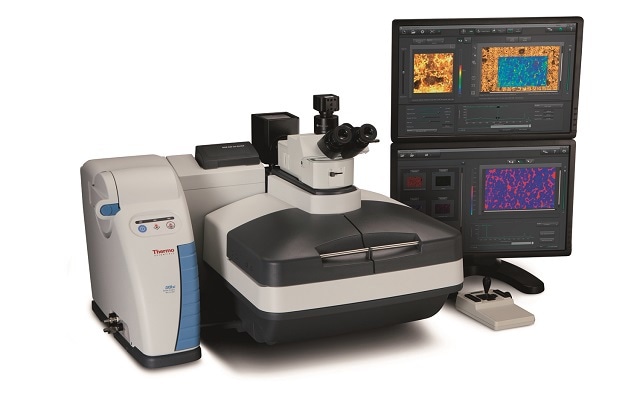
Figure 1. DXRxi Raman Imaging Microscope. Image Credit: Thermo Fisher Scientific - Vibrational Spectroscopy
Raman imaging can be used to elucidate the critical mechanisms and parameters governing nucleation and graphene growth via chemical vapor deposition (CVD). This knowledge underpins the development of scalable techniques suitable for engineering high-quality graphene samples that offer excellent properties.
This article from Thermo Scientific™ introduces the Thermo Scientific™ DXR™xi Raman imaging microscope, the latest tool for illuminating graphene growth via CVD synthesis.
Download the full article
Acknowledgments
Produced from materials originally authored by Thermo Fisher Scientific.

This information has been sourced, reviewed, and adapted from materials provided by Thermo Fisher Scientific - Vibrational Spectroscopy.
For more information on this source, please visit Thermo Fisher Scientific - Vibrational Spectroscopy.