In-situ scanning probe microscopy (SPM) imaging is provided by Hysitron on each of their nanomechanical testing platforms to yield superior nanomechanical characterization results and reliability.
In-situ SPM imaging provides high-resolution images at the point of the test by imaging with the same tip that is used to perform the nanomechanical testing. This provides crucial information concerning the surface topography of the test location and the resulting test deformation that would be unavailable without the image resolution offered by in-situ imaging.
Hysitron’s tip-scanning system allows imaging on any location of large samples or imaging of multiple samples without the need for user intervention.
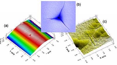
Figure 1. (a) Place an indent on the peak of an optical fiber (b) Measure cracks due to fracture (c) Place a scratch on the interface between two phases
Tip Placement
Precise positioning of the tip is required to reliably and repeatably obtain the desired information from nanomechanical tests. Whether testing multi-phase or single-phase materials, in-situ imaging is crucial to ensure that the test is placed at the desired location on the material.
In-situ imaging provides the ability to position the indenter tip with better than ten-nanometer resolution. This ultra-high precision positioning capability is often employed with multi-phase materials to allow selective characterization of each phase. Without the information provided by in-situ imaging, the user cannot be guaranteed that the test is placed within the desired phase.
Interfacial studies are virtually impossible without the precision of in-situ imaging, as placement of the indenter tip requires better resolution than what can be provided using motorized staging. Even homogenous materials typically possess surface features that must be avoided in order to eliminate testing anomalies due to features such as surface roughness.
Imaging of Deformation
In-situ imaging provides a wealth of information concerning the results of the test, in addition to the insight gained from the load/displacement data.
Gaining knowledge concerning the deformation incurred during the test allows insight into the material and the test results that may be unavailable without the SPM image.
Material property measurements, such as those for fracture toughness and yield strength, rely on information that can only be obtained from images that possess the resolution that this method can provide.
Subsequent SPM imaging may provide similar information if the site of the test can be found. However, the deformation that occurs on viscoelastic materials typically recovers with time, requiring that the time before imaging be minimized. In-situ images can be obtained seconds after completion of the testing, offering the most accurate representation of the real deformation. The recovery process can often be observed by repeated imaging of the test location, as the material flow allows some extent of recovery.
Observation of the mode and extent of recovery can play a significant role in providing the information necessary for future product development.
ScanningWear®
ScanningWear utilizes the synergy between the in-situ imaging and nanomechanical characterization to provide quantitative wear testing. By applying a load to the indenter tip as it is imaging the material surface, the surface is worn due to the applied stress under the tip.
This allows simulation of point contact or asperity wear and is suitable for a wide array of applications, from CMP to the paint industry. By obtaining images of the surface before and after the scanning wear test is performed, a wear volume can be quantified by calculating the volume of material removed.
A multitude of tip geometries are available to allow accurate simulation of the wear mechanisms in any application.
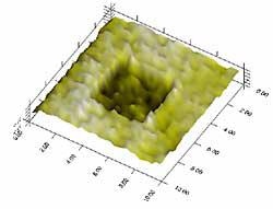
Figure 2. ScanningWear of a DLC Film
Image Analysis
Hysitron provides an exclusive offline image analysis software package, TriboView®, for quantitative analysis of in-situ SPM images. TriboView also provides advanced image viewing capabilities, such as 3D rendering, as seen in the above ScanningWear image.
TriboView also provides analysis options such as roughness analysis and section profiles, as demonstrated below on a PVD W film. The TriboView package has been designed to offer the user straightforward access to useful information for understanding and interpreting nanomechanical testing results.
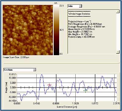
Figure 3. TriboView roughness analysis and section profile of II-01b an in-situ SPM image of a W film
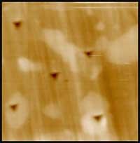
Figure 4. Selective testing of individual phases in a multi-phase material
Highlights
- Exclusive tip-scanning technique on Hysitron’s instruments.
- Provides tip placement within ten nanometers of desired test location.
- Eliminates blind testing of multi-phase materials or interfaces.
- Provides quantification of deformation features necessary for determination of material properties.
- Quantitative offline image analysis software.

This information has been sourced, reviewed and adapted from materials provided by Hysitron Incorporated.
For more information on this source, please visit Hysitron Incorporated.