Oxford Instruments’ Unity is a new detector for a groundbreaking new imaging technique in the Scanning Electron Microscope (SEM). It is the world's first Backscattered Electron and X-Ray (BEX) Imaging detector. It easily integrates backscattered electron and X-Ray signals to provide quick high-definition color images embedded with elemental data as the user navigates around the sample.
With a revolutionary design, which includes X-Ray and backscattered electron sensors inside the same sensor package, Unity is situated beneath the pole piece and has been designed to increase signal collection, which means that it can be employed at normal operating conditions and imaging speed, resulting in elementary imaging.
The high-speed BEX data collection of Unity makes it easier than ever before to image and analyze a whole sample in minutes, altering the way SEM is utilized.
From Instinct to Insight
Previously, examining a sample in an electron microscope has depended a lot on instinct. Normally, sample analysis starts with a greyscale image from a backscattered or secondary electron detector, moving around the sample, attempting to determine where the most productive regions for in-depth analysis are.
Acquisition settings are tuned to optimize settings for EDS analysis, and only following the EDS analysis will it likely determine if a user’s instinct was right. This process may be repeated several times before any useful results are generated, and it can take much longer to achieve a thorough comprehension of the sample.
Unity eliminates the guesswork by offering images instantly rich in useful elemental and topographic detail.
Why Unity?
The World’s First
Integrating two kinds of sensors inside one detector head, situated beneath the microscope pole-piece, the Unity detector gets its power and speed from its unique design.
Video Credit: Oxford Instruments NanoAnalysis
Dual Sensors
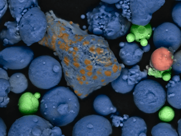
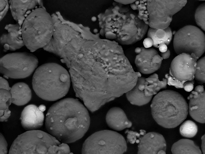
Similar to imaging detectors, Unity comprises backscattered electron (BSE) sensors that offer data about the sample density; these sensors reveal the different phases within a sample with a brightness associated directly with its atomic number.
Unlike standard imaging detectors, the X-Ray sensors gather characteristic X-Ray signals that deliver elemental information and enable Unity to instantly produce a color image. Signals from both types of sensor are layered to provide a sharp sample image that can be easily interpreted.
By instantly delivering such images, most guesswork and uncertainty are eliminated from the sample analysis, enabling microscopists to pilot their sample confidently and noticeably enhancing the analysis workflow.

Image Credit: Oxford Instruments NanoAnalysis
Designed for Daily Imaging
Alongside functioning at low beam currents, the Unity BEX imaging system offers X-Ray colored electron images throughout a broad array of operating settings, enabling users to utilize it in a similar manner to how they use a conventional imaging detector:
Flexible Working Distance
The new functioning position for the X-Ray sensors implies consistent chemical data can be gained throughout a broad array of Working Distances (WD).
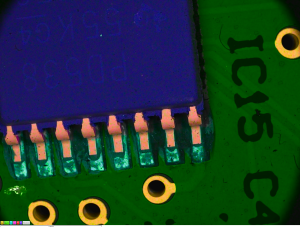
Image Credit: Oxford Instruments NanoAnalysis
Topography Is No Problem
The two Unity X-Ray sensors offer data from complex sample topography like deep troughs, which usually lead to no signal owing to shadowing. The two BSE sensors can be configured to topography mode, enabling the subsequent images to emphasize sample topography rather than sample density whenever required.
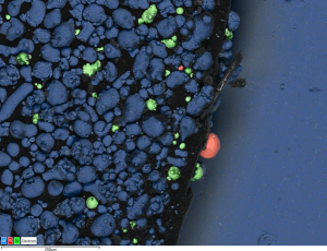
Image Credit: Oxford Instruments NanoAnalysis
Wide Field of View
Designed with a large aperture diameter, the Unity sensor head enables the microscopist to benefit from wide field modes—for example, fish-eye—to deliver whole sample analysis at the macro level and high-resolution imaging.
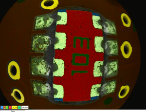
Image Credit: Oxford Instruments NanoAnalysis
Variable Pressure Mode
Allowing BEX imaging of non-conductive samples, Unity is built to function in variable pressure mode.
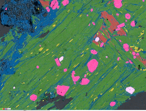
Image Credit: Oxford Instruments NanoAnalysis
High-Performance BSE Sensors
Different to traditional BSE detectors, Unity BSE sensors are custom shaped to increase signal collection and Peltier cooled for improved sensitivity, leading to a high-performance BSE detector.
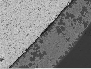
Image Credit: Oxford Instruments NanoAnalysis
Results Users Can Rely On
Unity relies on the technology and expertise built over several years of developing first-class X-Ray detectors. Combined with the AZtec software platform, the Unity detector is backed by high throughput X4 electronics, Tru-Q technology, and AZtec Live Chemical Imaging guaranteeing the sample imaging is fast and accurate.
Operating Position
Although BSE imaging detectors are usually located under the pole piece, X-Ray detection is typically only done with EDS detectors situated at a lower elevation angle and an increased distance from the sample. Placing the Unity detector head beneath the pole piece means the X-Ray sensors can take advantage of a much bigger collection angle and offer high signal at the same modest beam currents and dwell times utilized for BSE imaging.
With BEX imaging. Image Credit: Oxford Instruments NanoAnalysis
With traditional imaging. Image Credit: Oxford Instruments NanoAnalysis
Rapid Effortless Navigation
Large sample surface regions can be scanned and navigated for features of interest. As users move around, they see full-color images revealing the chemical components that make up the sample. As this detail is immediately visible, some examinations can be finished in minutes. Others can quickly move on to investigating more features or switch effortlessly to other techniques, like WDS, EDS, or EBSD, for more elaborate analysis.
The BEX technique's navigation speed allows Unity to revolutionize the typical microscopy workflow. Unity was shown to enhance microscope productivity up to 100 times in tests.
Video Credit: Oxford Instruments NanoAnalysis
Cartography
Unity imaging also facilitates precise, high-definition chemical mapping of the complete sample. What earlier might have consumed hours to acquire and likely needed an overnight run can now be gained in minutes.
In this video, Product Manager Haithem Mansour highlights the expansive area on the sample to be "mapped." To provide a sense of scale, the sample stub measures a total of 12 mm across. He then proceeds to select "Run" to initiate the acquisition process. In addition to the rapid pace of data gathering, Haithem emphasizes the remarkable quality of the map being created, as the software seamlessly stitches together individual images. It is extremely quick and easy to pick out small pink particles of the titanium contaminant despite the low level at which it is present.
Video Credit: Oxford Instruments NanoAnalysis
Unity in Action
It Is Time to Do Things Differently
Introduction to Unity BEX imaging detector
Introduction to Unity BEX Imaging Detector. Video Credit: Oxford Instruments NanoAnalysis