Jul 20 2015
An international research team, co-headed by Hans Elmlund, an associate professor from the Monash University’s ARC Centre of Excellence in Advanced Molecular Imaging, has devised a breakthrough imaging technique for capturing the 3D structures of nanocrystals, which find application in cancer treatment, pollution reduction, renewable energy collection.
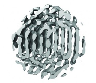 This a slab through the 3-D reconstruction of particle 1 along the vertical plane with tentative atomic positions indicated. ABC repeats of {111} planes are visible.
Scientists from Harvard, Boston, and Princeton Universities have also played a role in the development of this innovative technique, called “3D Structure Identification of Nanoparticles by Graphene Liquid Cell EM (SINGLE),” which enables the analysis of the 3D structures of these particles for the first time. Metallic nanoparticles have dimensions in the nanometer range, which makes it impossible to visualize their structure. This limitation has prevented researchers from gaining insights into how they work.
The study results have been reported in the Science journal. In this paper, the researchers have described the method in detail, along with its application in the characterization of the 3D structures of nanoparticles.
With the integration of three recently designed components, the novel imaging method delivers a performance far superior than earlier techniques. A graphene liquid cell is the first component, which is a one-molecule-thick bag capable of holding liquid within it during exposure to the ultra high vacuum of the electron microscope column.
A direct electron detector is the second component, which has a much higher sensitivity than conventional camera film. It is capable of capturing movies of the nanoparticle when they spin around in solution. A 3D modeling approach called PRIME is the final component, and this can generate 3D computer models of individual nanoparticles using the movies captured.
The movie clips, posted along with the publication, show the unprecedented details of the structure of two platinum nanoparticles. This in-depth information allowed the research team to gain new insights into the growth of these highly useful particles at individual atom level.
3D structure of nanocrystals.
Particle 1 in action | Monash University | Youtube.com
The field had expected cubical or at least highly symmetrical platinum nanocrystals. “It was surprising to learn that they form asymmetrical multi-domain structures,” Elmlund said.
Exploring how these nanoparticles form and evolve, and characterizing how they go through the transitions to achieve their final form are the next steps of the research team.
“It is important for us to understand this, so that we can design new materials, for example, to build better or more efficient solar cells, or make better and more economical use of fossil fuels,” Elmlund said.