Feb 1 2019
Cells in the human body have the potential to communicate with each other just like how people do. This interaction enables the organs in the human body to work in a synchronized manner, which consecutively allows humans to carry out a remarkable number of tasks encountered on a day-to-day basis.
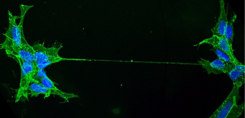
 Human neuronal cells connected by a nanotube. (Image credit: Anna Pepe/Institut Pasteur)
“Tunneling nanotubes”, or TNTs in short, are one of this means of communication. At the Institut Pasteur, a research team headed by Chiara Zurzolo used state-of-the-art imaging methods and eventually observed that the structure of these tunneling nanotubes challenged the very notion of cells. The results of the study have been reported in Nature Communications.
TNTs—as their name suggests—are very small tunnels that connect two or more cells and help in transporting a wide range of cargoes between them, such as viruses, ions, and the whole organelles. However, earlier studies performed by the same researchers (Membrane Traffic and Pathogenesis Unit) at the Institut Pasteur had demonstrated that TNTs play a critical role in the intercellular proliferation of pathogenic amyloid proteins associated with Parkinson’s and Alzheimer’s disease. This made scientists to suggest that TNTs act as an important avenue for the proliferation of neurodegenerative diseases in the brain and thus signify a new potential therapeutic target to halt the development of these incurable diseases. Furthermore, TNTs seem to have an active role to play in cancer resistance to treatment. However, researchers still do not know much about TNTs and the way they relate or vary from other kinds of cellular protrusions like filopodia, and hence, they opted to continue their studies to tackle these minute tubular connections in a more comprehensive manner.
The dogma of cell unit questioned
Therefore, a deeper insight into these minute tubular connections is needed because TNTs may have important implications in the health and diseases of human beings. Conversely, it has been very difficult to address this problem because of the fleeting and delicate nature of these structures, which do not endure traditional microscopic approaches. Hence, to resolve these barriers, scientists integrated a number of advanced electron microscopy techniques and subsequently imaged TNTs at temperatures below the freezing point.
Through this imaging technique, the scientists successfully interpreted the TNTs’ structure in greater detail. In particular, they demonstrated that the majority of TNTs—earlier shown to be single connections—are actually composed of several, smaller, separate tunneling nanotubes (iTNTs). The images of these nanotubes also revealed that there are thin wires that join iTNTs, which, in turn, could contribute to boosting their mechanical stability. Using time-lapse imaging, the researchers revealed the transport of organelles and thus demonstrated the iTNTs’ functionality. The team finally used a special kind of microscopy called “FIB-SEM” to create 3D images that have enough resolution to vividly detect that TNTs are “open” at either ends, and thus produce continuity between the two cells.
This discovery challenges the dogma of cells as individual units, showing that cells can open up to neighbors and exchange materials without a membrane barrier.
Chiara Zurzolo, Head, Membrane Traffic and Pathogenesis Unit, Institut Pasteur.
A new step in cell-to-cell communication decoding
By using an imaging work-flow that enhances upon, and prevents, earlier restrictions of tools used for analyzing TNTs’ anatomy, the investigators offer the first structural description of TNTs. Most significantly, they offer complete evidence that these are new cellular organelles with a distinct structure and are highly different from other known cell protrusions.
The description of the structure allows the understanding of the mechanisms involved in their formation and provides a better comprehension of their function in transferring material directly between (the cytosol of) two connected cells.
Chiara Zurzolo, Head, Membrane Traffic and Pathogenesis Unit, Institut Pasteur.
In addition, the team’s strategy, which conserves these fragile structures, will be handy for exploring the role of TNTs in other pathological and physiological conditions.
The study represents an important step towards deciphering cell-to-cell communication through TNTs and lays the foundation work for research into their physiological functions as well as their role in the proliferation of particles associated with bacteria, viruses, and other diseases, and misfolded proteins.
Les TNT remettent en question le concept de cellule /// TNTs challenges the very concept of cell
Video of Chiara Zurzolo’s team (in English), posted on Institut Pasteur Youtube channel. (Video credit: Institut Pasteur)