A Carbon nanotube (CNT), their name being derived from their size and composition, is a member of the fullerene structural family that forms cylindrical carbon molecules. A CNT can be up to several centimeters in length and its diameter is on the order of a few nanometers.
Properties of Carbon Nanotubes
CNTs are efficient conductors of heat and exhibit excellent strength and electrical properties. The extraordinary properties of CNTs make them suitable for a wide range of applications, such as optics, nano-electronics, materials and applications, to name a few. There are two main types of nanotubes and they inlcude single-walled (SWNTs) and multi-walled nanotubes (MWNTs). Other than impurities, CNTs are formed entirely of carbon atoms.
Structures of Carbon Nanotubes
The carbon’s structure is a hexagonal web rolled into a cylindrical tube, occasionally with a "cap" at either end of the cylinder (SWNT). Cylinders may also exist within other cylinders, similar to the layers of a leek vegetable, with 0.34nm gap between cylinders (MWNT). Additionally, some pentagonal (five-sided) and heptagonal (seven-sided) defects can cause cylinders to twist, bend, or change diameter.
Non-destructive Characterization of Carbon Nanotubes
Scanning tunneling microscopy or transmission electron microscopy is a proven characterization method for CNTs and provides the ability to measure the chiral angle or the diameter of a nanotube. However, the electron beam in the microscope could be possibly damage the nanotubes. Hence, for non-destructive characterization of the tubes, confocal Raman microscopy and atomic force microscopy (AFM) can be utilized. The Raman spectra of the tubes directly describe the molecular structure and chemical composition, while AFM provides information relating to the structural morphology.
Analysis of Carbon Nanotubes using Raman Microscopy
Depending on different electrical properties and polarization, it is possible to identify the diameter of the tubes, their orientation, and even the differentiation between SWNT and MWNT. To show the capabilities of confocal Raman microscopy, this study examines CNTs using the WITec alpha300 R system.
Wavelength Dependent Raman Imaging
Raman signals of CNTs depend on the wavelength of the excitation laser. To this end, Raman spectroscopy on SWNT is a resonant Raman scattering process of single photons and provides a better understanding on the electronic and photonic structure of one-dimensional carbon molecules in their bonding environment.
The radial breathing modes (RBM), which can be found at low wave numbers (120-350/cm), are caused by coherent vibration of carbon atoms in the radial direction. The RBM’s position provides information on the nanotube’s diameter; the peak’s intensity reveals the electronic structure of individual tubes and the combination of both parameters enables the characterization of the SWNTs’ structure.
Raman Scattering
Similar to all resonant processes, the intensity of the Raman scattering from SWNT depends on the wavelength of the excitation laser. This study demonstrates results from one and the same sample area investigated with 514nm (Figure 1a and b) and 633nm (Figure 1 c and d) excitation wavelength, respectively.
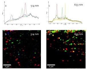
Figure 1. Confocal Raman imaging on CNTs. (a) Typical spectra at 514nm; (b) Distribution of corresponding nanotubes on the silicon substrate; (c) Typical spectra at 633nm; distribution of corresponding nanotubes.
SWNTs with a diameter ranging from 0.9nm (Figure 1a red spectrum) to 1.42nm (Figure 1a blue spectrum) are detected using a 514 nm excitation. The distribution of SWNTs is color coded in Figure 1b. Using a 633nm excitation laser, the SWNTs are identified with structural properties meeting the resonance conditions.
Applications of Carbon Nanotubes
Biomedical Field
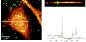
Figure 2. (a) Confocal Raman imaging of CNTs on a cell; (b) Depth scan along the white line indicated in Figure 2a; (c) Spectra used for generating the images in Figures 2a and 2b.
For potential applications of CNTs in the biomedical field, it is vital to have a thorough knowledge about the interaction of CNTs with individual cells. In this study, CNTs were applied to single cells in a cell culture and imaged using confocal Raman microscopy subsequent to fixing on the substrate.
Figure 2a depicts the distribution of CNTs (green) on the cell. To establish the level of uptake of the nanotubes in the cell, a depth scan was carried out in x-z plane along the indicated line. Figure 2b displays the distribution of the nanotubes in the cell, clearly showing that the nanotubes are located both on the cell surface and within the cell. Figure 2c displays the spectra utilized for producing the images. The green spectrum denotes the CNTs, while the red spectrum relates to the cell.
Microelectronic Devices
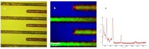
Figure 3. a) Video image of electrodes of an integrated circuit. (b) Raman image of the electrodes showing an accumulation of the nanotubes near the electrodes. (c) Spectrum of the Si-Substrate and a nanotube at the electrode.
Since CNTS exhibit diverse electronic properties, several attempts have been made to integrate then into circuits for improved circuit design. For instance, confocal Raman Imaging can be utilized to detect the nanotubes with regard to electrodes and other interconnects. In addition, the properties of the nanotubes can be determined thoroughly by their spectra.
In this experiment, confocal Raman microscopy was used to image the electrodes of a silicon integrated circuit. Figure 3a shows the microscopic video image of the circuit, which does not display any CNTs, while the Raman image in Figure 3b shows that the nanotubes (red) accumulate close to the electrodes. AFM in a combined system configuration of the alpha300 series can be used to determine whether the nanotubes are coupled to the electrode or not. Figure 3c displays a spectrum of such a nanotube showing that the nanotube is a semiconducting type.
Conclusion
WITec’s confocal Raman microscope alpha300 R can be used to gain a better understanding of the physical and mechanical properties of CNTs. WITec develops high-performance instrumentation for both scientific and industrial applications.

This information has been sourced, reviewed and adapted from materials provided by WITec GmbH.
For more information on this source, please visit WITec GmbH.