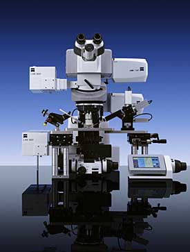
Zeiss has overcome the challenges of imaging deep inside living tissues with the launch of the LSM 7 MP, a purpose-built multiphoton laser scanning microscope that, for the first time, incorporates two separate scanners. The twin scanners mean that the compact system's two excitation lasers can be set to different wavelengths and used either simultaneously or sequentially for specimen imaging and manipulation.

With the scan module of the LSM 7 MP optimised for excitation light up to 1100 nm, efficient fluorescence excitation deep inside tissue samples is possible without the phototoxic damage associated with high intensity light. Highly sensitive non-descanned detectors or a unique non-descanned GaAsP detector with signal outcoupling directly above the objective lens ensure vivid, high resolution fluorescent imaging, even in whole, live animal studies.
Application fields include high resolution 3D imaging in long-term observations of development processes and functional imaging in conjunction with simultaneous photo-manipulation. In combination with the Axio Examiner upright microscope stand and the AxioCam camera, the LSM 7 MP is an optimal system for performing highly specialized multiphoton microscopy applications. A wide range of detectors, filters and other accessories allows individual users to configure a personalised and application-specific system. Quick set-up and ease of use are guaranteed thanks to the intuitive ZEN imaging and control software.
Conventional confocal microscopes quickly lose their capacity to resolve fluorescent structures in thicker specimens due to the absorption and scattering of both the excitation and emitted light. In the multiphoton LSM 7 MP, fluorescence is only excited if at least two photons are absorbed by a fluorochrome molecule within less than a femtosecond (10–15 seconds) and the whole of the emitted light signal can be used for imaging.