The SEM Visualization Engine (SEM-VE) combines a 16 bit scan generator and dual super-sampling signal acquisition hardware with image processing and control software for your Zeiss electron microscope. The SEM-VE system enables acquisition of images up to 32k x 32k pixels (one gigapixel image size), with dwell times from 100 ns to > 100 s, adjustable in 100 ns increments. Images may be saved with eight or sixteen bits of intensity. The SEM-VE "Mosaic Tool" is designed to create large image montages and is highly integrated with SmartSEM.
The Mosaic Tool enables the automated acquisition of multi-image Mosaics at multiple sites on the sample, automatically moving from "image Tile" to Tile, and Mosaic site to site, executing SmartSEM autofunctions such as Focus, Stigmation, Brightness, etc. as required. The end result is an "Extreme Field of View" image montage that can cover optical microscope scale (or even naked eye scale) areas of your sample, at SEM nanometer scale resolution.
Given suitable samples, unattended operation can be performed over a period of days, automatically acquiring Terabytes of image data at rates up to 30 Gigabytes of image data per hour. This allows the user to collect data from a given region at a resolution approximately seven orders of magnitude smaller than the region. For example it is possible to acquire a 40 square millimeter region, at 4 nanometer resolution, in under 4 days.
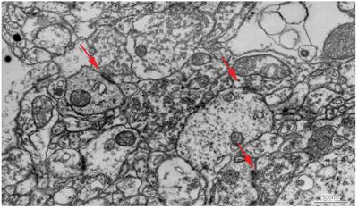
Single 30 nm thick section of mouse brain imaged with backscattered electrons at 8 kV using a Zeiss Sigma FE-SEM. Original image resolution was 4 nm per pixel. Red arrows point to areas where one neuron contacts and transfers information to another.
Extreme field of view image montages improve the complete understanding of structures by providing detailed image information from the macro scale to the nanoscale.
Need for Large Images
Why acquire "EXTREMELY LARGE" images?
- Reduces the number of tiles to acquire, reducing stage motion delay and more importantly reducing the areal fraction of each image "lost" to overlap. For a 4 nm pixel size and a 2 µm stage accuracy, overlap can be 66% of the image area at 3k x 2k (33% new data), but only 6% of the image area at 32k x 32k (94% new data / image) thus a 2.5 mm x 2.5 mm mosaic at 4 nm resolution would require ~92,000 images with a conventional frame store, but only 400 images when using the SEM-VE.
- Reduces the number of overlap "seams", leading to less beam damage and degradation of the sample.
- These factors greatly reduce computational complexity - considerably easier to stitch and align when working with 400 images rather than 92,000. This reduction in computational complexity becomes even more critical when three dimensional alignment of serial sections is required.
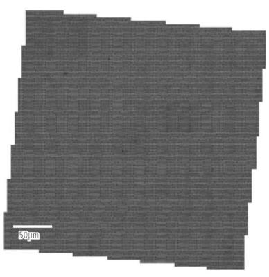
Image tile boundaries are hidden.
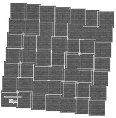
The position of image tile boundaries are displayed (white lines)
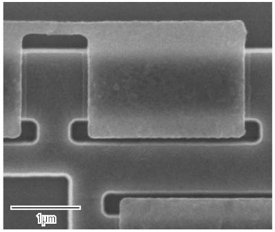
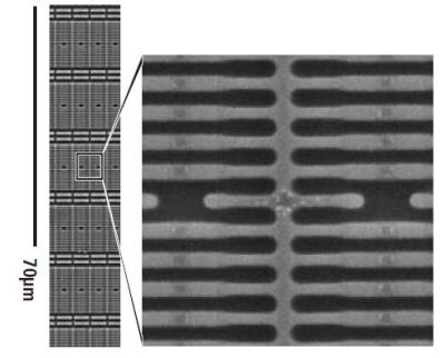
A 65 nm technology node graphics processor integrated circuit, stripped to its silicon substrate with HF acid etching.
The mosaic consists of 49 images, each ~500 Megapixels, automatically stitched by the VE-Viewer into a ~1/3 mm x 1/3 mm mosaic. This mosaic was acquired stepping at an angle, with scan rotation used to align image features to the scan axes, in order to accentuate the stitching for illustrative purposes.
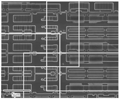
A magnified view of the junction between four tiles as shown above, with tile boundaries displayed in white, as otherwise the boundaries between tiles are virtually seamless.
A sample of the high resolution detail visible across the entire Extreme Field of View mosaic.
XFOV - STEM in SEM
By using the GEMINI® Multi-mode STEM detection system, the information limit for the SUPRA™ FE-SEM can be extended beyond the nanometer range. A resolution of 0.8 nm is now readily attainable and gives additional nano scale information. The quality of the image obtained with the STEM unit are similar to images obtained by a TEM with a scanning attachment.
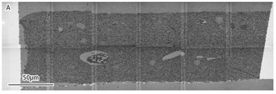
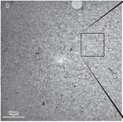
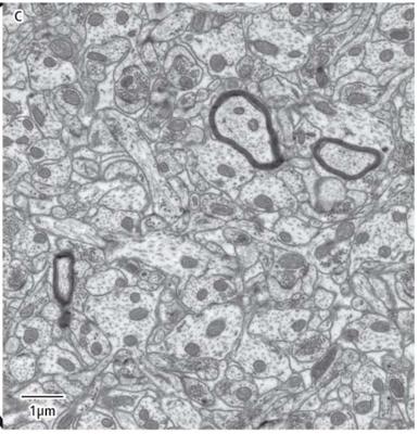
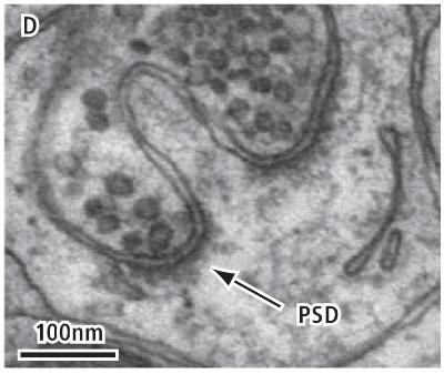
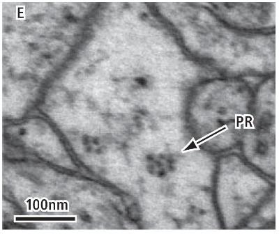
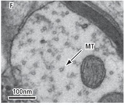
STEM-in-SEM images acquired on a Zeiss SUPRA™ 40 equipped with SEM-VE. Images show a single 45 nm thick post-section stained epoxy ultra section of perfused rat hippocampus. A series of images were automatically acquired from multiple sites on multiple TEM grids. (A) Low magnification overview image of a single mosaic site consisting of 6 x 2 tiles, each 49 µm x 49 µm, acquired using the GEMINI® STEM detection system in a SUPRA™ FE-SEM. Each image tile was acquired at 2 nm per pixel. (B) A single 49 µm SEM-in-STEM image, acquired at 2 nm per pixel. The inset square illustrates the comparative size of a TEM image acquired on a 4k x 4k camera, also at 2 nm per pixel. Acquiring larger fields of view reduces both distortion and the requirements for image stitching. (C) Detail from an approximately 9 µm x 9 µm region of Image B.
Note the image quality is comparable to results obtained by conventional TEM. At full resolution, key organelles are readily resolved including (D) post synaptic densities at synapses, (E) polyribosomes and (F) cross-sectioned microtubules suitable for serial tracing and dense reconstruction.
Advantages of SEM-VE
How Does SEM-VE Help to Solve Your Problems
- Acquire images up to 1 Gigapixel in a single scan, increased from previous ~7 Megapixel maximum.
- Continuously selectable scan size from 1 x 1 to 32k x 32k pixels.
- Continuously definable field of view and pixel resolution.
- Scan rates selectable from 100 ns to >>100 s dwell per pixel, in 100 ns increments.
- Save image data as 8 or 16 bit TIFF files.
- Automation enables unattended operation, with automatic stage motion, focus, stigmation, brightness and contrast adjustment as required.
- Acquire and stitch high resolution image Mosaics at optical fields of view with nanometer resolution.
- Includes the VE-Viewer, an image viewer tailored to efficiently handle the multi-gigabyte Mosaics produced by SEM-VE. The VE-Viewer allows the user to open, stitch, navigate and intelligently re-render the large 2D datasets produced by SEM-VE.
A high magnification excerpt from the long image.
A long, narrow single image approximately 1/3 mm x 20 µm in size acquired at 10 nm per pixel resolution and 200 ns dwell time. This image took approximately 15 seconds to acquire.
SEM-VE is available on new and currently installed instruments as a field upgrade.
.jpg)
This information has been sourced, reviewed and adapted from materials provided by Carl Zeiss.
For more information on this source, please visit Carl Zeiss.