The importance of studying bone at the nanoscale
Developing an effective and robust system for deepening our understanding of the characteristics of the tissue, structure, mechanical properties and organization of bone at the level of the nanoscale, as well as for expanding our understanding of bone interactions at this scale, is imperative for advancing medical techniques for treating bone-related disorders.
Gaining this scale of information would allow researchers to further medical understanding of characteristics such as bone strength, stiffness, and toughness, which are all factors that impact the way healthcare professionals assess and treat aged and diseased bones. This information would also be fundamental in the development of new treatments for osteoporosis, as well as for the development of the bio-inspired materials of the future. For example, there is currently a thriving sector for medical science dedicated to innovating new and improved bone-like materials for various applications, and the closer these materials can be made to mimic the real thing, the better they will perform.
Recent years have seen a rapid growth of interest in the use of x-ray diffraction techniques as a non-invasive, non-destructive method for analyzing the nanostructure of bone to help advance our knowledge of human bone, having significant implications for the future of healthcare.
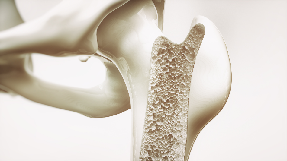
Image Credit: Crevis/Shutterstock.com
What is x-ray diffraction?
X-ray diffraction is a well-established method for identifying structural properties, texture and orientation pattern, crystalline phases, crystal size, crystallinity, and perfection. The emittance of an x-ray beam onto a sample interacts with the photon matter of the target material. An electron in an outer shell of the sample becomes excited at the location where the x-ray beam hits it. This displaces the electron, creating a hole in the element’s electronic structure. Following this, an electron from a higher energy shell moves in to replace the ejected electron, and the resultant energy released from this action as an x-ray is picked up by a detector. Measuring the amount of energy released is indicative of many features of the material. It is this information that helps characterize the sample.
Why use x-ray diffraction to characterize the nanocomposites of bone?
Bone has a structural hierarchy at different length scales, making it a unique substance and therefore difficult to copy, which has implications on the area of medical science that is attempting to create materials mimicking human bone. Scientists recognize that it is vital to understand the structure, organization, characteristics of the underlying structural components, and nature of interactions at the nanoscale level.
X-ray diffraction reveals how macroscopic bone characteristics are impacted by microstructural behavior, something that is not achievable with other techniques. It can be used to reveal details of bone at the nanoscale, allowing scientists to gain a thorough understanding of the properties of bone that have remained elusive due to the limitations of previously used techniques that have only been able to gain information on the bulk behavior of bone.
Bone encompasses the unique characteristics in stiffness, strength, weight, and fracture toughness, which is governed by the hierarchical structure from the nanoscale to the macroscopic scale. Previous studies have already described the basic structure of bone, down to the mesostructure level (0.5–10 mm). More recently, x-ray diffraction methods have been successfully implemented to reveal the hierarchical structure of bone at the nano-scale.
Research using this technique has uncovered that it is made up of an organic phase (32–44% bone volume), an inorganic phase (33–43% bone volume) and water (15–25% bone volume). X-ray diffraction has also revealed that type I collagen is the man component of the organic phase and that apatite-like mineral crystals, such as hydroxyapatite, are the main elements of the inorganic phase.
X-ray diffraction studies have also demonstrated how the two-phase composites of bone, collagen and apatite minerals, interact to give bone its characteristic of strength. Previously the details of this interaction had eluded scientists, but with x-ray diffraction, it has been discovered that there is a mineral matrix with collagen inclusions which contribute to strength, as well as water also playing a factor in this characteristic.
X-ray diffraction has effectively been used in studies of strain measurement of bone. In these studies, the crystalline planes are used as strain gauges. Families of crystallographic planes within a sample of bone diffract x-rays in distinct ways dependent on the amount of strain they are under, allowing scientists to understand the behavior of bone at the nano-scale while under certain amounts of strain. Further to this, x-ray diffraction techniques have uncovered even more about the nature of strains on bone through measuring the impact of different external loads on mineral crystals in all orientations.
Finally, x-ray diffraction has been used to investigate the deformation of bone at the nanoscale level. Through numerous landmark studies over the years, significant insights have been gained into how mineral crystals in bone deform under different external loads, as well as on the fundamentals of lattice deformation, profile broadening, and texture.
Osteoporosis
Osteoporosis is common in the elderly population and can lead to deterioration of both mental and physical health, lowered a person’s quality of life. Gaining a deeper understanding of the characteristics of human bone at the nanoscale opens the potential for developing new and more effective treatments and preventions.
Mounting evidence that has been building over the years has demonstrated that x-ray diffraction is a highly useful method in gaining insights on the structural and mechanical characteristics of bone at the nanoscale, furthering our knowledge of how bone form, degrades, and heals, advising the development of better bone-related treatments, as well as advancing the development of the bio-inspired materials.
Sources:
Londoño-Restrepo, S., Jeronimo-Cruz, R., Millán-Malo, B., Rivera-Muñoz, E. and Rodriguez-García, M. (2019). Effect of the Nano Crystal Size on the X-ray Diffraction Patterns of Biogenic Hydroxyapatite from Human, Bovine, and Porcine Bones. Scientific Reports, 9(1). https://www.nature.com/articles/s41598-019-42269-9
Müller, F. (2018). Bone-inspired Biomaterials. Bioinspired Materials Science and Engineering, pp.337-350. https://onlinelibrary.wiley.com/doi/10.1002/9781119390350.ch17
Tadano, S. and Giri, B. (2011). X-ray diffraction as a promising tool to characterize bone nanocomposites. Science and Technology of Advanced Materials, 12(6), p.064708. https://iopscience.iop.org/article/10.1088/1468-6996/12/6/064708/meta
Disclaimer: The views expressed here are those of the author expressed in their private capacity and do not necessarily represent the views of AZoM.com Limited T/A AZoNetwork the owner and operator of this website. This disclaimer forms part of the Terms and conditions of use of this website.