Sep 29 2009
Instead of the classic scalpel, surgeons can also operate with an electroscalpel. A significant advantage to this technique is that while a cut is being made, blood vessels are closed off and hemorrhaging eliminated. Now another advantage may be added as well: a German-Hungarian research team has developed a mass-spectrometry-based technique by which tissues can be analyzed during a surgical procedure. As the team led by Zoltán Takáts reports in the journal Angewandte Chemie, it may be possible to distinguish between malignant tumor cells and the surrounding healthy tissue in real time during cancer surgery. Until now, precise histological examination of the removed tissue has followed after tumor surgery, and has required several days. If it reveals that the tumor has not been completely removed, a second operation is needed. The new method may spare patients this second surgery in the future.
 © Wiley-VCH
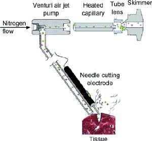
In electrosurgery, tissue is locally exposed to high-frequency electrical current in order to guide a cut, remove tissue, or halt bleeding. The tissue being treated becomes very hot and is partially vaporized. The electrical current also generates electrically charged molecules during the vaporization. The team of scientists from the University of Giessen, the Budapest firm Massprom, Semmelweis University, and the National Research Institute for Radiobiology and Radiohygiene, also in Budapest, made use of this process for their new method called rapid evaporation ionization mass spectrometry, or REIMS. They equipped an electrosurgical instrument with a special pump that sucks the vaporized cell components up through a tube and introduces the charged molecules into a mass spectrometer.
It turns out that mainly lipids, the components of cell membranes, are registered by the mass spectrometer. “Different tissue types demonstrate characteristic differences in their lipid composition,” explains Takáts. “Tumor tissue also differs from healthy tissue.” The scientists were able to develop a special algorithm to unambiguously identify and differentiate between types of tissue.
“Tissue analysis with REIMS, including data analysis, requires only fractions of a second,” according to Takáts. “During an operation, the surgeon thus received virtually real-time information about the nature of the tissue as he was cutting it.” This opens new vistas for cancer surgery in particular: the method helps to precisely localize the tumor during surgery and to delimit it from the surrounding healthy tissue. REIMS also provides information about whether the carcinoma is in an early or advanced stage.