The VivaTome for fluorescence microscopes from Carl Zeiss is an optimal tool for developmental and cell biologists to examine the dynamics of living specimens without extensive prior know- ledge of optical sectioning. For the visualization of fast processes, VivaTome is an attractively-priced solution that offers outstanding ease of use and excellent results. It is designed for applications where temporal resolution is a priority. This system provides clear, easy-to-interpret and quantifiable results for biological specimens-regardless of whether it is a cell structure, a tissue section or a living organism.
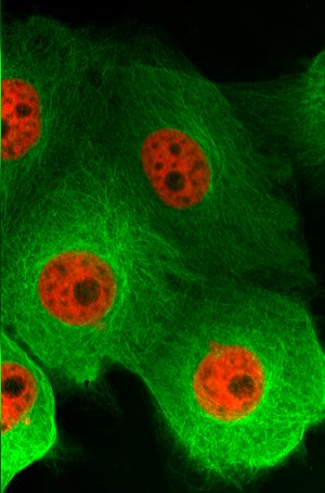 Epithelial cells of a pig’s kidney (LLC-PK1), marked with Tubulin-Emerald and H2B-mCherry, photographed with the VivaTome microscope module and the 63x/1.4 Plan-APOCHROMAT objective lens from Carl Zeiss.
With a low level of technical complexity, VivaTome delivers high-quality microscope images that have only been possible with highly advanced systems until now. Using simple and economical white light illumination, frame rates of up to 30 frames a second are achieved. With the VivaTome, the entire spectrum of ZEISS objective lenses can be used with a consistently high optical section quality.
VivaTome captures the widefield and the optical section data simultaneously, ensuring that no information is lost. It can be attached to the ZEISS Axio Observer, Axio Imager and Axio Examiner microscope systems using a simple c-mount. Existing widefield systems are easily retrofitted.
The VivaTome is the result of the collaboration between Carl Zeiss and Aurox Ltd. (Oxford, UK). It is based on the aperture correlation principle developed by Tony Wilson and collaborators at the University of Oxford.