Jan 30 2012
An international team of scientists has for the first time crystallised a key enzyme of the pathogen for African sleeping sickness in a living cell and investigated it with the world's strongest X-ray laser.
This new method will open new ways to study the structure of biomolecules, the scientists write in the professional journal "Nature Methods". In the case of African sleeping sickness that affects more than 60 million people mainly in Central Africa this method might lead to a new treatment approach against the parasite Trypanosoma brucei.
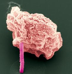
 Electron microscope image of an insect cell with a protein crystal sticking out. Credit: Michael Duszenko/Universität Tübingen.
The three-dimensional structure of a biomolecule gives biologists clues about its function, and in the case of a pathogen it also offers the perspective to block a harmful protein with a tailor-made artificial molecule. For example, if the enzyme cathepsin B of Trypanosoma brucei is blocked, the parasite will die. However, the structure analysis of biomolecules is a difficult and time-consuming process. Normally, a sufficiently large crystal of the protein in question has to be grown in the lab before it can be investigated with X-ray light of a synchrotron radiation source.
Crystal growing is complicated and often takes weeks or even months. Therefore, the team of scientists - among them scientists from the universities of Tübingen, Hamburg and Lübeck, and from Deutsches Elektronen-Synchrotron DESY in Hamburg - chose another approach. With the help of a virus, they inserted the genetic blueprint for cathepsin B into living insect cells. The infected cells started to produce the enzyme incessantly, and with the steadily increasing concentration, the enzyme eventually crystallised. After about 70 hours, the micrometre-small crystals became visible in the microscope, some of them even sticking out of the cells.
At the US accelerator centre SLAC in California, the scientists bombarded these crystals with the world's strongest X-ray free-electron laser LCLS. Although its intensive X-ray flash completely vaporises the crystals in less than a billionth of a second, it is bright enough to previously take a detailed diffraction image of the crystal, making it possible to calculate the structure of the crystallised enzyme. However, to gain the complete structural information the experiment must be repeated very often with a large number of crystals, which was not part of the study.
But the result shows that with the new technology it is possible to generate high-quality data of the protein structure of nanocrystals. "Our experiments have shown that the promise of X-ray lasers to revolutionize structural biology is indeed becoming true," said DESY scientist Prof. Henry Chapman from the Center for Free-Electron Laser Science (CFEL). "We have shown that previous limitations to protein crystallography can be overcome by using pulses of X-rays so intense, that they transform the proteins into a dense plasma similar to the conditions inside the sun. Yet the pulses are so short that fine details are seen before destroying the sample," Dr. Anton Barty from CFEL added.
Apart from the Federal Ministry of Education and Research-funded young investigators group "Structural Infection Biology Using new Radiation Sources (SIAS)" of the universities of Hamburg and Lübeck, and the Hamburg School for Structure and Dynamics in Infection (SDI) of the State of Hamburg Excellence Initiative, the research was done with the participation of a team of scientists headed by Professor Michael Duszenko from the University of Tübingen, a CFEL-group headed by Professor Henry Chapman as well as other DESY scientists and international collaborators. CFEL is a cooperation of DESY, the Max Planck Society and the University of Hamburg.
"Our result shows that the super lasers offer completely new possibilities for the structure determination of biological macromolecules, and perhaps the days will be over soon when we needed months or even years to grow crystals of certain proteins being large enough for X-ray radiation sources at synchrotrons" said SIAS leader Dr. Lars Redecke, one of the main authors of this study.
As from 2010, SIAS - an initiative of the structural research scientists Professor Christian Betzel, University of Hamburg, and Professor Rolf Hilgenfeld, University of Lübeck - investigates the use of innovative radiation sources for structural determination of proteins and other biological molecules.
The European X-ray laser European XFEL, currently being built in Hamburg, will open a unique opportunity for biomolecule investigation. Already today, DESY operates the free-electron laser FLASH for soft X-ray radiation.