Feb 13 2014
Many of the most striking recent observations in biology have been made using super-resolution microscopy techniques.
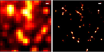 A diffraction-limited microscopy image (left) and a super-resolution microscopy image processed with rainSTORM (right) of vesicles labelled with fluorescent Epidermal Growth Factor
These allow scientists to look at objects that are smaller than the diffraction limit of light (around 250 nanometres), which is a fundamental limit of traditional optical microscopes.
The NPL and Cambridge research concerns a super-resolution technique called localisation microscopy, which uses fluorescent labelling and computer modelling to bypass the diffraction limit. The highlighted paper summarises the methods used to process the captured images and provides a series of MATLAB scripts known collectively as rainSTORM. This software takes raw data and performs image analysis and visualisation, and is freely available to anyone who is interested.
Localisation microscopy works by using fluorescent dyes to label molecules within cells. These dyes can be switched on and off, and by doing so the centre position of each individual fluorescent molecule can be identified and plotted. This plotted position is known as a 'localisation'. The process is repeated thousands of times to build up a highly detailed, super-resolution image using all of the plotted positions from each repetition. Software is a vital part of this technique.
Alex Knight, who worked on the research at NPL, said:
"This research demonstrates the importance of choosing the right software and using it properly. The combination of the paper and the freely available rainSTORM scripts will hopefully help spread best practice in super-resolution microscopy techniques, eliminating many of the problems faced when interpreting the results."
By allowing scientists to identify fine structures within cells and to study them in more detail, localisation microscopy, and other super-resolution techniques, could give new insights into the smallest features of cells and improve how we understand and treat diseases. NPL is working to develop complementary super-resolution techniques to improve both their temporal and spatial resolutions, thereby increasing their effectivenss and enabling scientists to study ever smaller structures.