Jul 14 2016
Carbon nanotubes attract a lot of interest in diverse fields such as nanomechanics, photonics, electronics, and quantum optics, as they can be manufactured in a number of properties and shapes, and used in numerous applications. It is important for researchers to have an instrument that could establish these properties in a fast and precise manner.
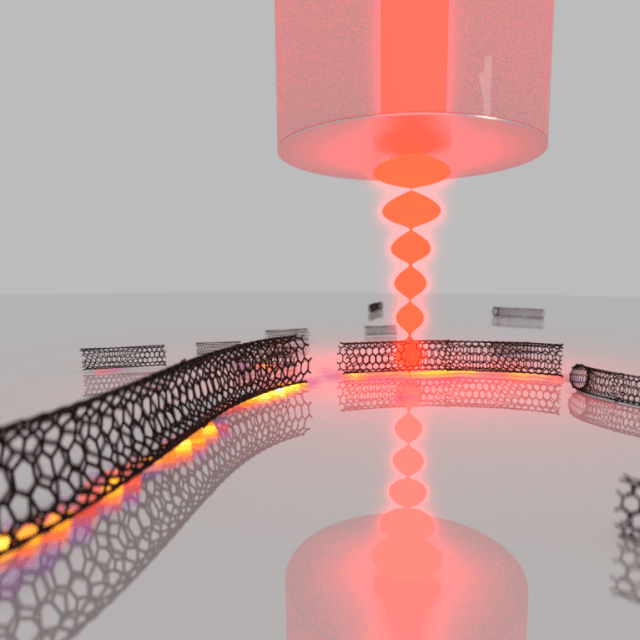 Schematic illustration of the experiment. (Graphic: MPQ, Laser Spectroscopy Division)
On the whole, Raman spectroscopy is receptive to the chemical structure that facilitates rise to these properties. But the signals are fundamentally weak and therefore require optimization methods.
Recently, researchers from the Laser Spectroscopy Division of Prof. Theodor W. Hänsch (Director at the Max Planck Institute of Quantum Optics and Chair of Experimental Physics at the Ludwig-Maximilians-Universität, Munich) have formulated a method to improve Raman scattering signals using an optical microcavity, which then is used for molecular diagnostics by integrated Raman and absorption imaging.
In contrast to other methods, the new technique just relies on increased vacuum fluctuations of the electromagnetic field within a cavity, which provides a significant enhancement without unnecessary background, making the method a feasible tool for molecular imaging.
Each molecular species possesses its own fingerprint of vibrational frequencies, which conveys data regarding its chemical structure. Raman spectroscopy enables optical detection of the vibrational spectrum in a powerful manner by inelastic light scattering. As an optical method, it can enable spatial imaging and integrate high spatial resolution with chemical contrast. This capability creates potential for a number of applications for Raman microscopy, spanning from characterization of nanomaterials and the analysis of biological samples to industrial process monitoring.
The current research focuses on analyzing individual carbon nanotubes. Nanotubes are available in a wide range of diameters and can either be semiconducting or metallic. Raman spectroscopy is mostly sensitive to the molecular structure that monitors these properties, and Raman imaging enables determining this for separate nanotubes. However, the standard Raman scattering encounters inherently low signal, which is very bad for imaging applications and when examining individual nanosystems.
Our approach is to place the sample of nanotubes, dispersed on a substrate, inside of a microscopic cavity, where optical resonances can be harnessed to enhance the Raman scattering process. At the same time, the cavity can be scanned across the sample and focusses the light to a spot size not too far from the diffraction limit, such that high resolution images can be generated. The cavity amplifies both the Raman scattering process as well as absorption from the sample. This allows one to combine ultrasensitive absorption microscopy with Raman imaging within a single measurement.
Dr. David Hunger, Scientist, Max Planck Institute of Quantum Optics
To make the cavity enrichment effect large, small cavities capable of storing light for several thousands of circulations are needed – which is quite a challenge when scanning capabilities for imaging operations are also required.
In the microcavity installation constructed by Dr. David Hunger and his team, one side of the resonator is composed of a plane mirror that acts simultaneously as a carrier for the sample under evaluation. The counterpart is a powerfully curved micro mirror on the end facet of an optical fiber. Through this fiber, laser light is coupled into the resonator.
With regard to the fiber, the plane mirror is moved point by point so as to bring the sample step by step into the focal point of the cavity mode. Simultaneously, the distance between both mirrors is modified such that the cavity’s resonance condition is matched with a resonance of a Raman scattering technique. Positioning accuracy in the range of tens of picometers is required for this to be achieved.
To obtain a full Raman spectrum, we step-wise tune the mirror separation to sweep a cavity resonance across the desired spectral range and collect the cavity-enhanced Raman scattering signal. Since the cavity resonances are extremely narrow, this can lead to a spectral resolution way beyond the capabilities of conventional Raman spectrometers.
Thomas Hümmer, PhD Student, Mac Planck Institute of Quantum Optics
The Raman signal is greatly improved because of the Purcell effect, which is obtained from the intensifying vacuum fluctuations and the prolonged lifetime of the photon inside the microcavity. In the experiment, this results in improving the resonant light by up to a factor 320. A more than 6-fold increase is obtained by the cavity experiment when the net signal received from a single Raman line from the cavity is compared to the signal received with the most appropriate standard microscope. Additional improvements in the future will help to enhance this development by several orders of magnitude.
The complete potential of the method is then illustrated by cavity-enhanced hyperspectral imaging. In this type of a measurement, cavity-enhanced Raman spectra are noted at several locations on the mirror, and a spatial image can be created, showing, for example the line shape or the strength of Raman lines.
In our experiment we study one particular Raman transition, which is sensitive to the diameter and the electronic properties of the nanotube. From the hyperspectral image we can deduce the size of a large set of individual tubes and determine whether they are metallic or semiconducting.
Thomas Hümmer, PhD Student, Mac Planck Institute of Quantum Optics
The technique's applicability to a large number of samples makes it a potential tool for single molecule Raman imaging. Additionally, the system could be adapted to construct Raman lasers with an array of novel materials, or it might be applied to obtain quantum control over molecular vibrations.