May 24 2019
Surgical medical meshes were invented about five decades ago and since then have become important elements in the recovery processes of damaged-tissue surgeries, with hernia repair being the most frequent one.
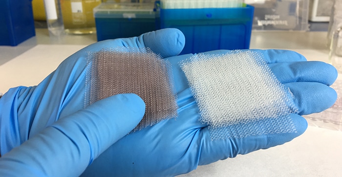 Surgical implants, covered with gold nanoparticles (pile of meshes on the left) compared to the original surgical meshes previous to the treatment (pile of meshes on the right). (Image credit: ICFO)
When these meshes are implanted inside the patient’s tissue, the conformable and flexible design of these products helps in holding the muscles tightly and enables patients to recover more quickly when compared to the traditional sowing and stitching surgery.
Conversely, whenever a medical implant is inserted into a patient’s body, bacterial contamination can potentially occur at the time of the surgery and this would subsequently lead to the formation of an infectious biofilm over the surgical mesh surface. Such kinds of biofilms can behave similar to an impermeable coating and prevent any kind of antibiotic agent from reaching and destroying the bacteria developed on the biofilm and eventually stop the infection.
Therefore, antibiotic therapies, which are time-restricted, are likely to fail against these highly resistant bacteria and the patient can end up in repeated surgeries that might even result in death. In fact, in 2015, over 30,000 deaths in Europe have been associated with infections caused by antibiotic-resistant bacteria, according to the European Antimicrobial Resistance Surveillance Network (EARS-Net).
Earlier, a number of methods have been pursued to prevent contamination of implants at the time of the surgery. To combat these antibiotic-resistant bacteria, post-surgery aseptic protocols have been recognized and implemented but still, none was able to overcome this problem.
In a new study reported in Nano Letters and emphasized in Nature Photonics, ICFO researchers have developed an innovative method that utilizes photonics and nanotechnology to considerably enhance the performance of medical meshes for surgical implants. The study involved ICFO team Dr Ignacio de Miguel and Arantxa Albornoz, headed by Romain Quidant, ICREA Professor at ICFO, in association with scientists Irene Prieto, Dr Christine Weis, Dr Pau Turon, and Dr Vanesa Sanz from B. Braun—the leading medical device and pharmaceutical device company.
Since 2012, the researchers at B. Braun Surgical, S.A. and ICFO have been collaborating continuously and now they have created a novel medical mesh with a specific feature—the mesh surface was chemically altered to anchor a countless number of gold nanoparticles. This was done because gold nanoparticles can efficiently change light into heat at highly localized regions.
In earlier studies, the method of utilizing gold nanoparticles in light-heat conversion processes had already been verified in the treatments for cancer. In addition, at ICFO, this method had been applied to a number of earlier studies, which were supported by the Cellex Foundation. This is yet another prominent example of how previous visionary philanthropic support, which was addressed at dealing with important issues, ultimately paves the way to major practical applications. For this specific case, when the researchers came to know that over 20 million hernia repair operations are performed every year worldwide, they assumed that this technique can possibly lower the medical costs in recurrent surgeries and, at the same time, eliminate the ineffective and costly antibiotic treatments that are presently being used to address this concern.
Therefore, in their new in-vitro experiment and through a comprehensive process, the researchers used millions of gold nanoparticles to coat the surgical mesh, evenly distributing them over the whole structure. They also examined the medical meshes to guarantee the non-degradation of the material, long-term stability of the particles, and the release or non-detachment of nanoparticles within the surrounding environment (flask). Then, with the help of a scanning electron microscope, the team observed a uniform distribution of the nanoparticles across the structure.
After the customized mesh was ready, the researchers exposed it to S.aureus bacteria for a period of 24 hours until they viewed the development of a biofilm on the mesh surface. Later, they subjected the medical mesh to short powerful pulses of near-infrared light (800 nm) for about 30 seconds to make sure that thermal equilibrium was achieved, and then repeated this treatment for 20 times with rest intervals of 4 seconds between every pulse. The researchers discovered the following factors:
Firstly, they observed that illuminating the medical mesh at the particular frequency would promote localized surface plasmon resonances in the gold nanoparticles—a mode that causes the efficient conversion of light into heat, while burning the microorganisms at the surface. Secondly, when the researchers used a fluorescence confocal microscope, they were able to observe the amount of bacteria that still alive or had died. For the microorganisms that continue dot be alive, they noticed that the biofilm bacteria were turned into planktonic cells, recovering their weakness or sensitivity towards antibiotic treatment and also to immune system response. With regards to the dead bacteria, the researchers observed that when the amount of light delivered to the mesh surface is increased, this causes the bacteria to lose their adherence and peel off the surface of the mesh. Thirdly, the researchers confirmed that operating at near-infrared light ranges was fully compatible with in-vivo settings, which means that a technique like this is not likely to cause damage to the surrounding healthy tissue. Finally, the team repeated the treatment and demonstrated that the repeated heating of the medical mesh did not have an effect on its conversion efficiency capabilities.
The results of this study have paved the way towards using plasmon nanotechnologies to prevent the formation of bacterial biofilm at the surface of surgical implants. There are still several issues that need to be addressed but it is important to emphasize that such a technique will indeed signify a radical change in operation procedures and further patient post recovery.
Romain Quidant, ICREA Professor, ICFO
Our commitment to help healthcare professionals to avoid hospital related infections pushes us to develop new strategies to fight bacteria and biofilms. Additionally, the research team is exploring to extend such technology to other sectors where biofilms must be avoided.
Dr Pau Turon, Director, Research and Development, B. Braun Surgical, S.A.